



Alveolar Bone Grafting
Restoring Continuity of the Upper Jaw and Dental Arch
What Is Alveolar Bone Grafting?
What Patients Find Most Helpful To Know About Alveolar Bone Grafting

Supports Permanent Tooth Growth
The graft creates the bone needed for adult teeth to erupt properly.

Timing Is Important
Surgery is typically performed before the canine tooth erupts to ensure proper dental alignment.

Bone Integrates Naturally
The BMP or transplanted bone gradually fuses with the surrounding jawbone as healing occurs.

Orthodontic Treatment Is Often Involved
Braces or other dental treatments may be used before and after surgery to guide tooth movement.
When Is Alveolar Bone Grafting Performed?


Improved support for permanent teeth

Restoration of the dental arch structure

Better stability for orthodontic treatment


Reduced communication between the mouth and nose

Improved gum health and dental function

Provides support for future dental care

Alveolar Bone Graft Procedure
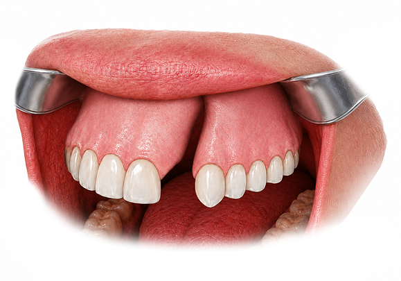
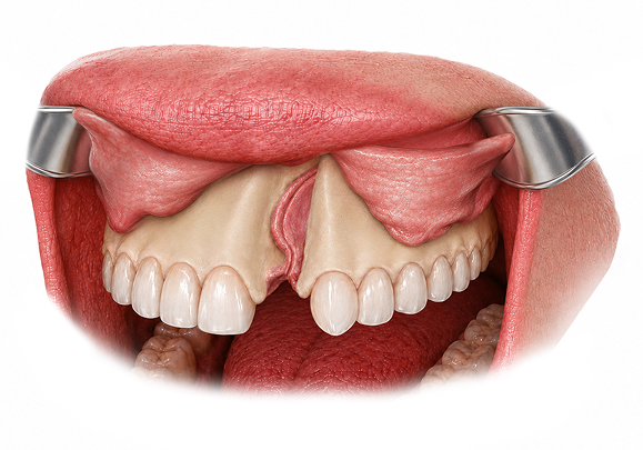
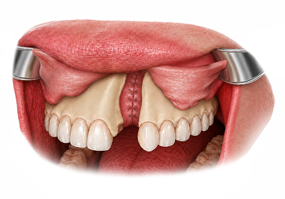
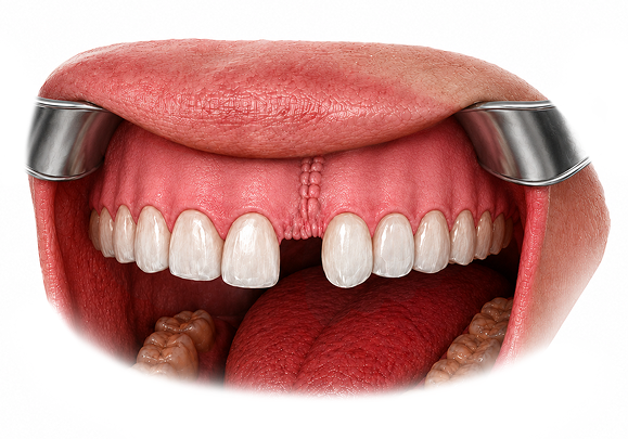
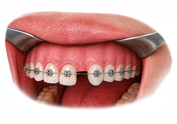
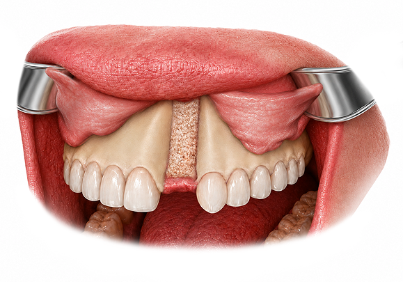
During surgery, the tissues surrounding the cleft are carefully elevated to expose the defect. The nasal floor is then repaired to separate the mouth from the nasal cavity and create a stable foundation for the graft. Bone morphogenetic protein (BMP), or in rare cases bone from the hip, is placed within the cleft, and the gum tissues are closed over the graft. As healing occurs, new bone forms within the cleft, creating support for tooth eruption and future orthodontic treatment.
Before & After Gallery
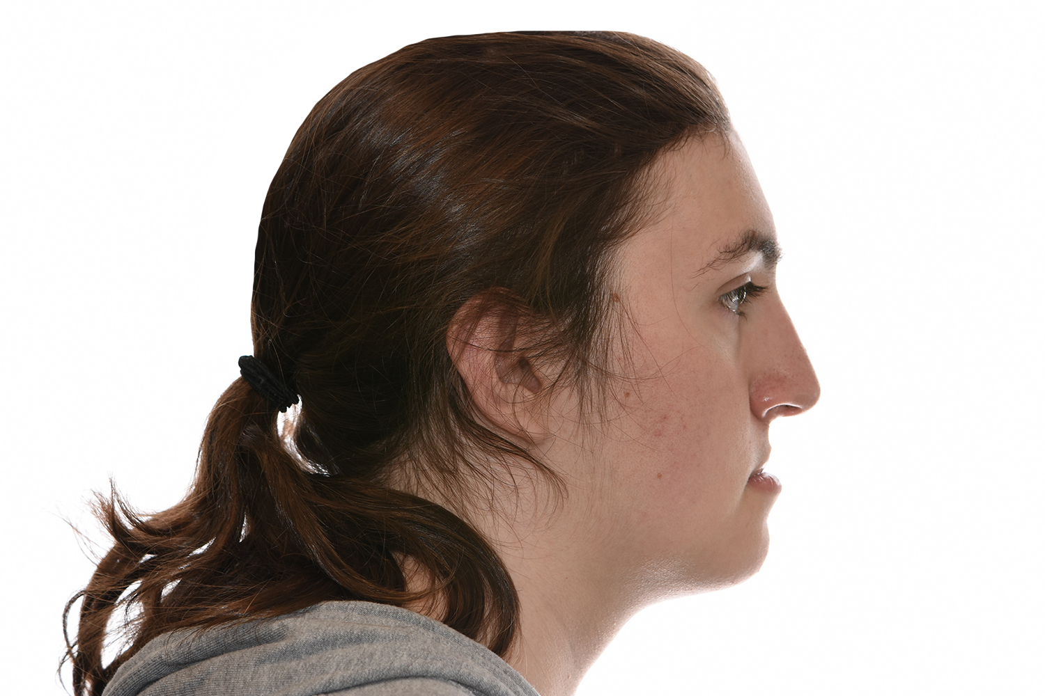
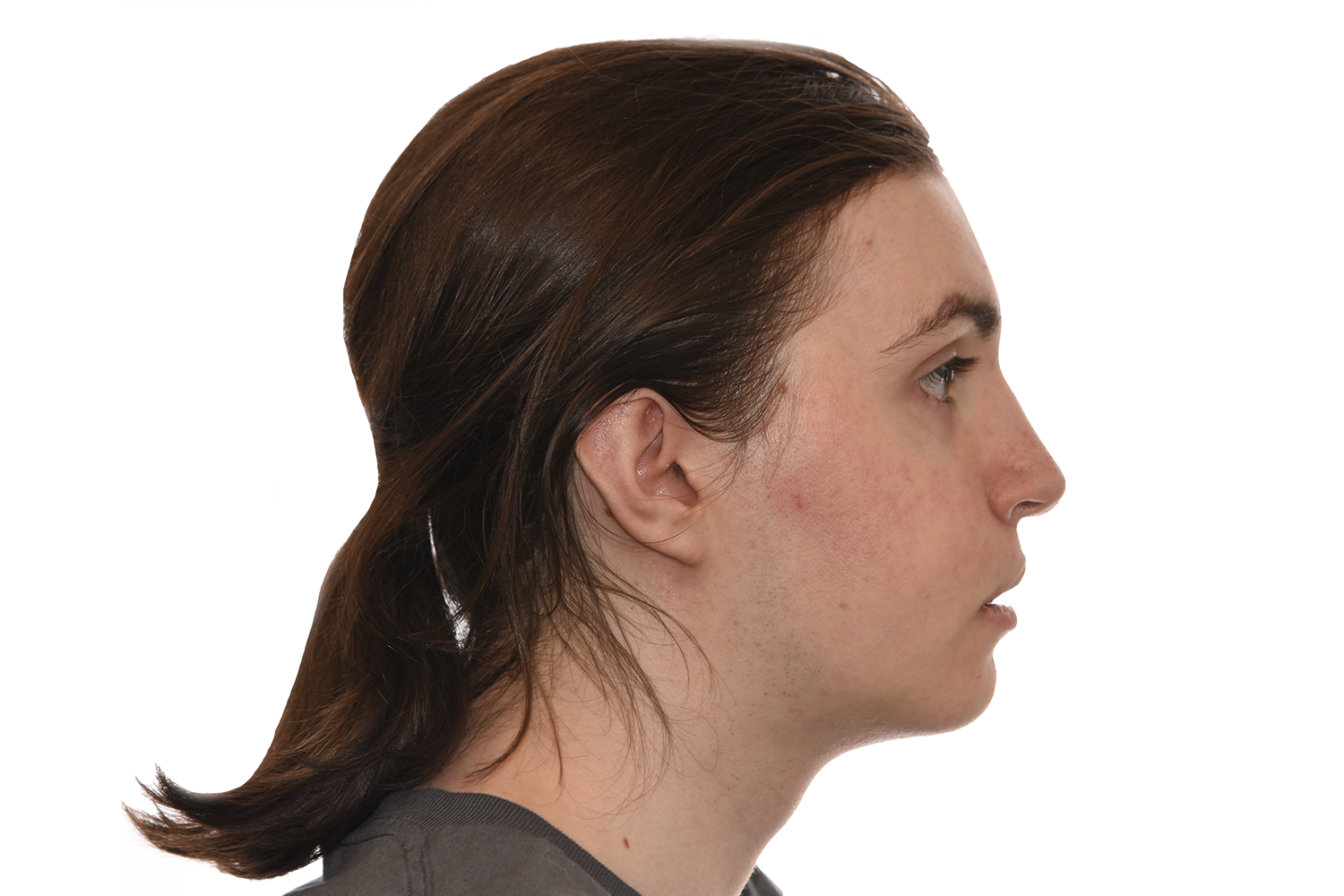
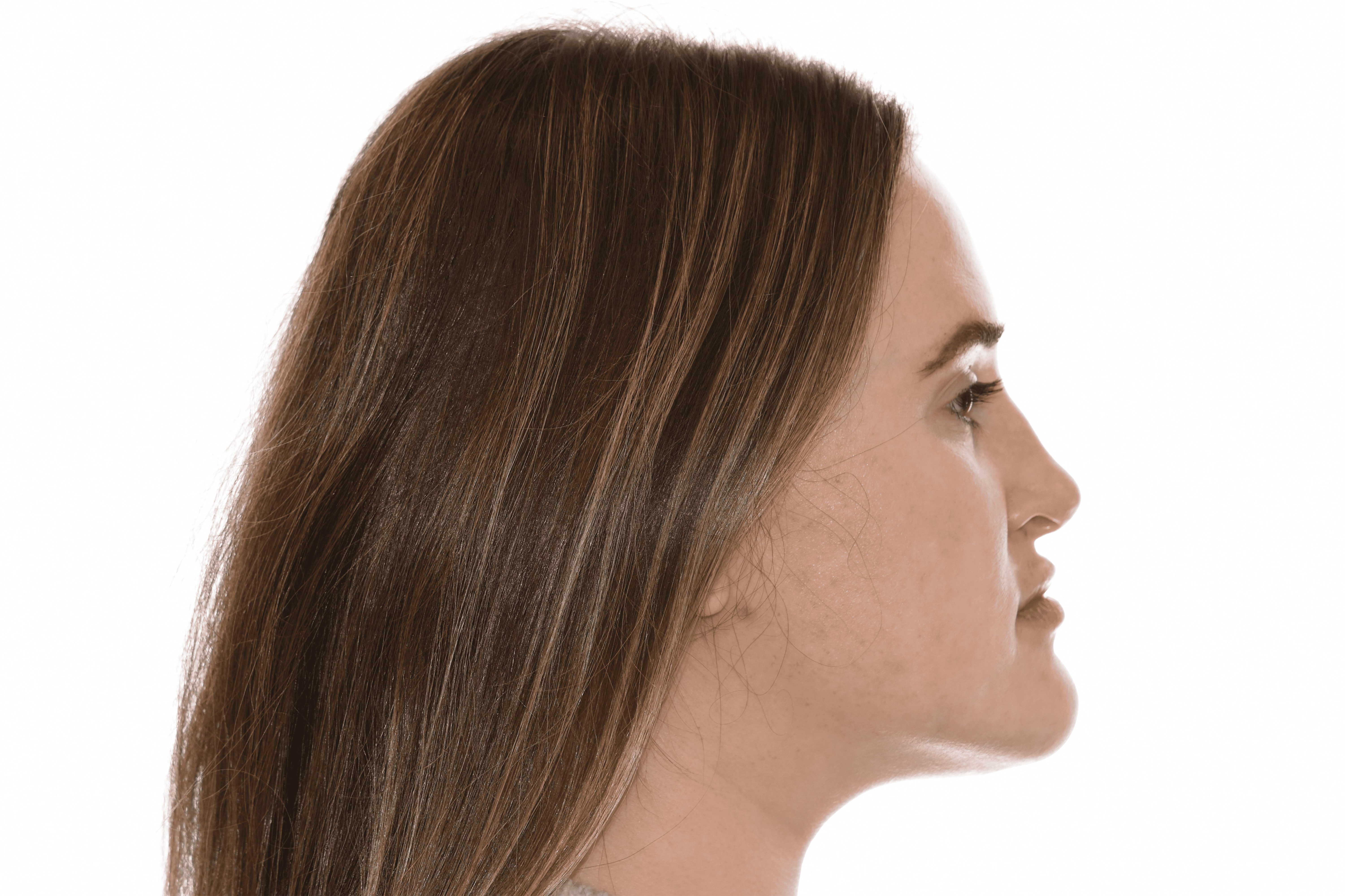
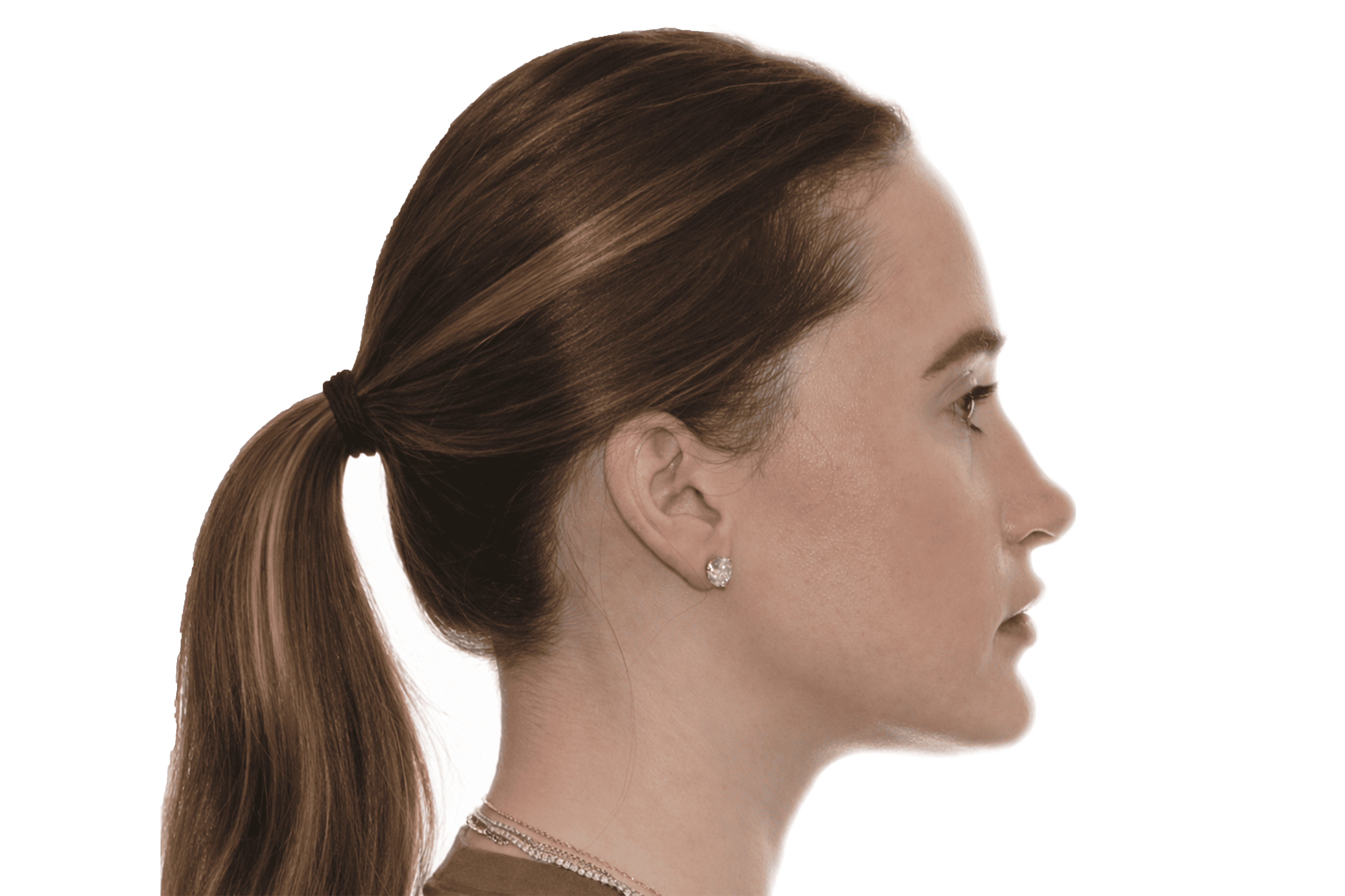
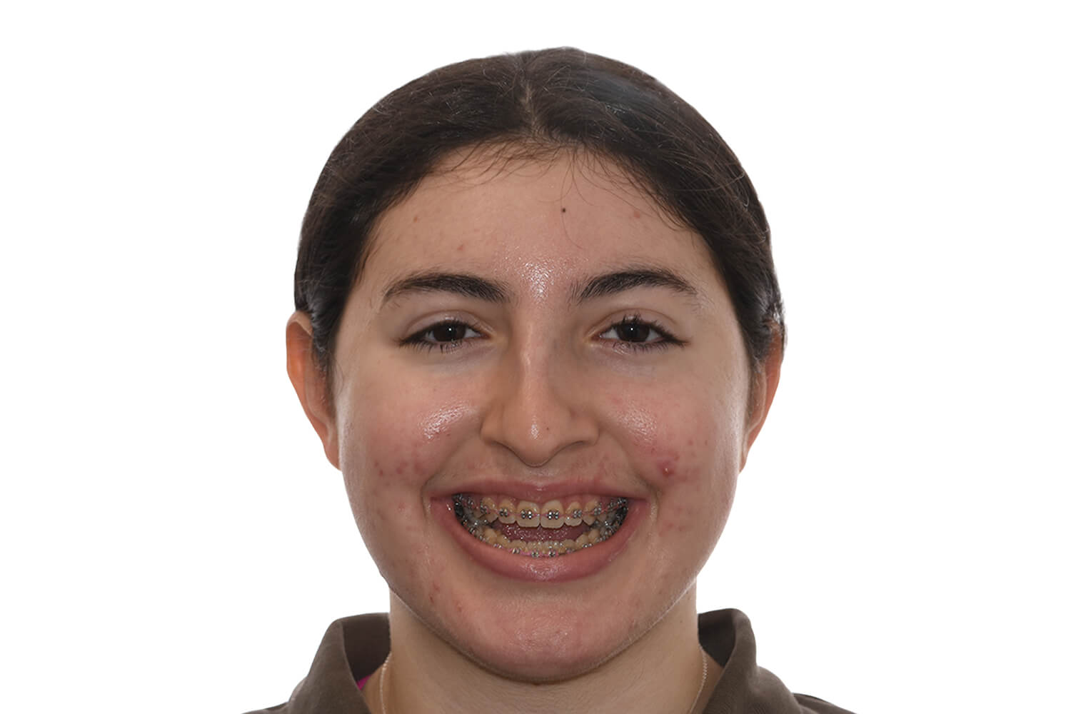
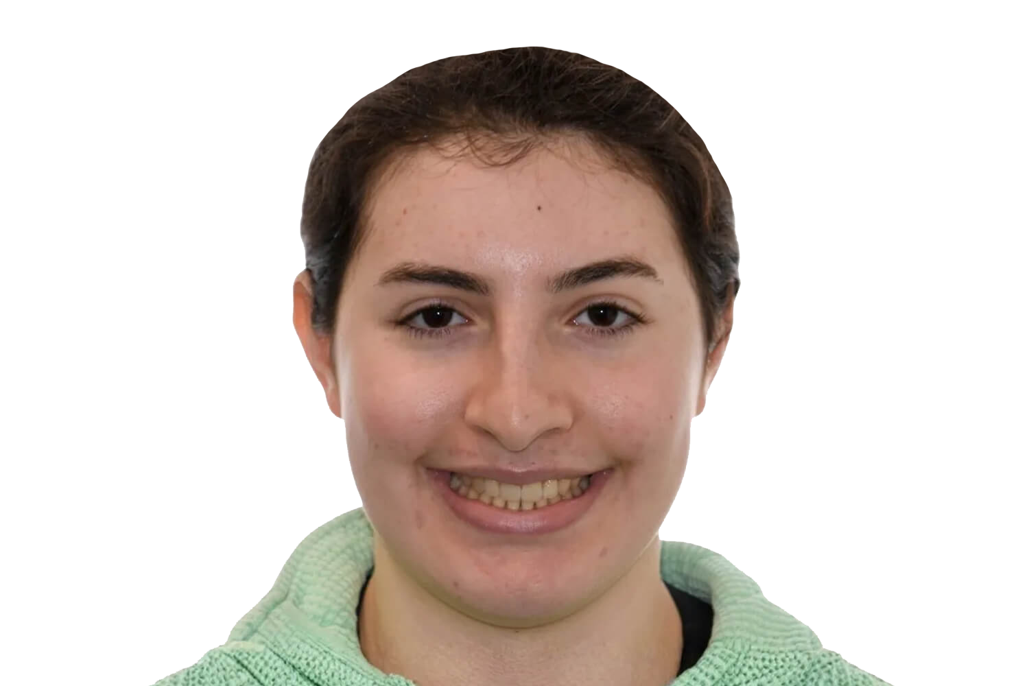
Timeline For Alveolar Bone Grafting
Recovery
Following alveolar bone grafting, recovery focuses on protecting the graft site, supporting healing, and allowing bone to integrate predictably.
First Week
Early Healing
Week 2–4
Initial Recovery
Month 1–3
Bone Integration
Month 3–12
Long-Term Results
What to Expect After Alveolar Bone Grafting
Most children recover well and return to normal activities within 1–2 weeks.
Risks & Possible Complications

An experienced team and close follow-up care help maximize the likelihood of optimal outcomes.
Alveolar Bone Graft requires precise surgical judgment, specialized training, and familiarity with the anatomical and functional nuances of this procedure. Outcomes depend heavily on the surgeon's experience, technique, and ability to anticipate and manage the full range of possible findings. Surgeons who perform this procedure regularly are typically better positioned to achieve consistent, safe results.
In a private practice setting, care is often more streamlined and consistent, with the same surgeon closely involved in evaluation, treatment planning, surgery, and follow-up.
In academic or hospital-based settings, care may involve a larger team that can include residents or fellows participating under supervision. Both models can provide excellent care, but the structure and continuity of treatment may feel different. At our center, your care is delivered directly by your surgeon — not by residents, fellows, or surgeons in training.
It is important to understand who is performing each part of your procedure. In some settings, the attending surgeon performs all critical portions, while in others trainees may assist or participate under supervision. This should be discussed clearly in advance. At our center, your surgeon performs all critical portions of the procedure.
Depending on the treatment setting, residents, fellows, or students may be involved in aspects of care. Patients should feel comfortable asking about their role and how care is supervised.
At our center, there are no residents or fellows involved in performing surgery.
Post-operative follow-up is an important part of surgical care. At our center, your surgeon personally follows you through recovery and any additional visits required.
At our center, your surgeon is directly involved at every stage — from the initial consultation and treatment planning through surgery, post-operative visits, and long-term follow-up.
Alveolar bone grafting is a procedure that fills the bony gap in the upper gum ridge — the alveolus — that is present in most children born with cleft lip and palate. The graft restores the continuity of the dental arch, provides a bony foundation for erupting permanent teeth, and closes any persistent communication between the mouth and nose.
Grafting is most commonly performed during the mixed dentition period, typically between ages seven and ten, timed to the eruption of the permanent canine tooth adjacent to the cleft. Orthodontic expansion of the upper arch is required before surgery to align the bone segments and prepare the site for grafting.
Two primary graft approaches are used. The traditional approach uses the patient's own bone harvested from the hip — specifically the iliac crest — which is considered the gold standard for graft integration. An alternative approach uses bone morphogenetic protein, or BMP, combined with a carrier material to stimulate bone formation without a donor site harvest. Both approaches have good evidence supporting their effectiveness, and the choice is made based on the patient's anatomy, the size of the defect, and other clinical factors.
Hip bone grafting provides the patient's own living bone cells and is associated with excellent long-term integration. It requires an additional incision at the hip for bone harvest, which adds some discomfort and a small scar at the donor site. BMP eliminates the donor site entirely, reducing post-operative discomfort and the need for a second surgical site, but it is a more costly option and may not be appropriate for all defect sizes and patient profiles. Both options are discussed with families during the consultation.
Yes. Upper arch expansion and alignment are typically completed before bone grafting to ensure the bone segments are in the correct position to receive the graft. Close coordination between the orthodontist and surgeon is essential to achieving a good result.
The procedure is performed under general anesthesia. When using hip bone, the surgeon makes a small incision at the hip to harvest cancellous bone, which is then packed into the alveolar cleft through an incision in the gum tissue. When using BMP, the carrier material containing the protein is placed directly into the cleft without a donor site. The graft site is then closed with sutures.
For hip bone grafting, most patients are admitted for one night due to the donor site. For BMP grafting, the procedure is typically performed on an outpatient basis with same-day discharge. Both approaches are performed under general anesthesia.
A soft diet is required for several weeks while the graft heals. Hip graft patients experience some donor site soreness at the hip for one to two weeks, which is managed with medication and resolves with time. Most children return to school and light activity within approximately one to two weeks. Follow-up imaging is performed after several months to confirm graft integration and monitor tooth eruption.
Yes. We regularly care for families traveling from outside the region for alveolar bone grafting.
For hip bone grafting, most patients are discharged after one overnight stay. Out-of-town families should plan to remain locally for approximately one week primarily to attend their first post-operative visit, at which the surgeon examines the graft site and donor site, confirms healing, and clears the family for travel home. For BMP grafting, the procedure is outpatient and patients are discharged the same day. Out-of-town families undergoing BMP grafting should still plan for approximately one week locally for the post-operative visit, as confirmation of early healing is important before travel. The specific timeline may vary based on the child's age and recovery progress, and your surgeon will confirm the post-op visit schedule in advance.
Yes. Once the post-operative visit has taken place and healing is confirmed, most families are cleared for ground travel shortly thereafter. Air travel is generally appropriate at approximately seven to ten days after surgery. For hip graft patients, donor site comfort is also a consideration for longer travel, and your surgeon will advise on this at the post-op visit.
Yes. Follow-up imaging at several months after surgery is required to confirm graft integration and monitor tooth eruption through the grafted site. Continued orthodontic treatment follows grafting, and the grafted area is monitored over time. Some follow-up may be conducted via telemedicine while imaging and in-person assessment visits will require a return to the center.
The consultation includes clinical and imaging assessment of the alveolar cleft, review of the child's dental development, coordination with the orthodontist to confirm readiness for surgery, and a detailed discussion of the graft material options, the procedure, recovery expectations, and follow-up plan.