



Maxillary Distraction
Controlled, Gradual Advancement of the Upper Jaw for Greater Precision and Long-Term Stability
What Is Maxillary Distraction?
What Patients Find Most Helpful to Know About Maxillary Distraction

At-Home Distraction
At-home distraction typically becomes routine after the first day or two.

Distraction Arms Get Removed
Distraction arms are removed after activation, which is usually completed around week three.

First Week Is the Toughest
Healing becomes noticeably easier after the initial 7–10 days.

Jaws Usually Aren’t Wired Shut
The jaws are not wired shut after surgery or during the distraction process.
When Is Maxillary Distraction Performed?


More predictable correction of upper jaw underdevelopment

Improved bite, chewing efficiency, and speech

Enhanced facial balance and midface projection


Gradual adaptation of surrounding soft tissues for safer and more predictable results

Long-term skeletal stability and more predictable outcomes

Reduced risk of relapse compared to single-step advancement
Before & After Gallery
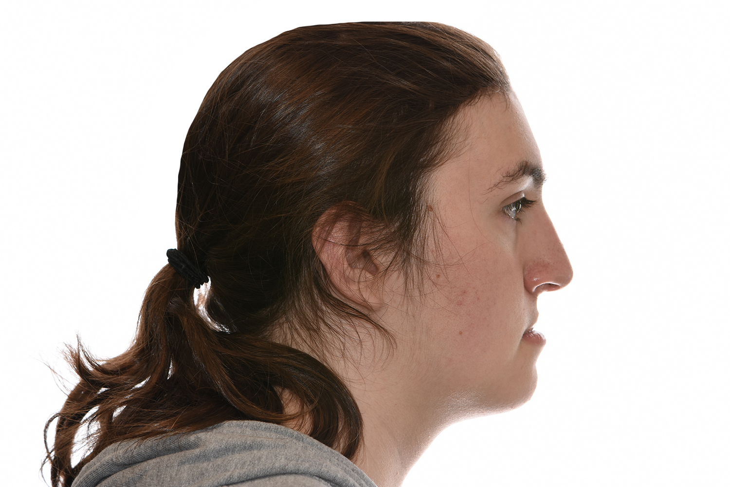
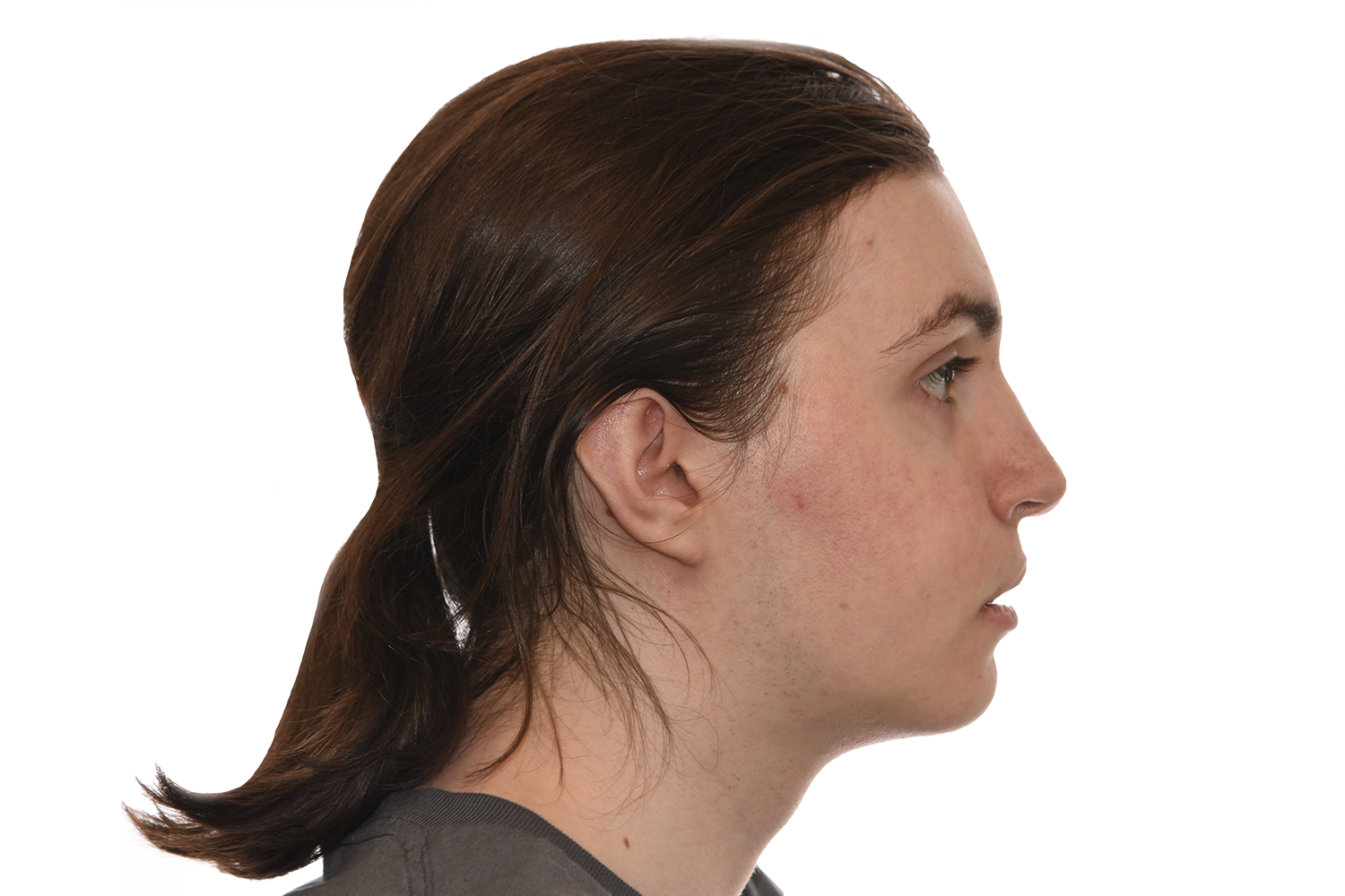
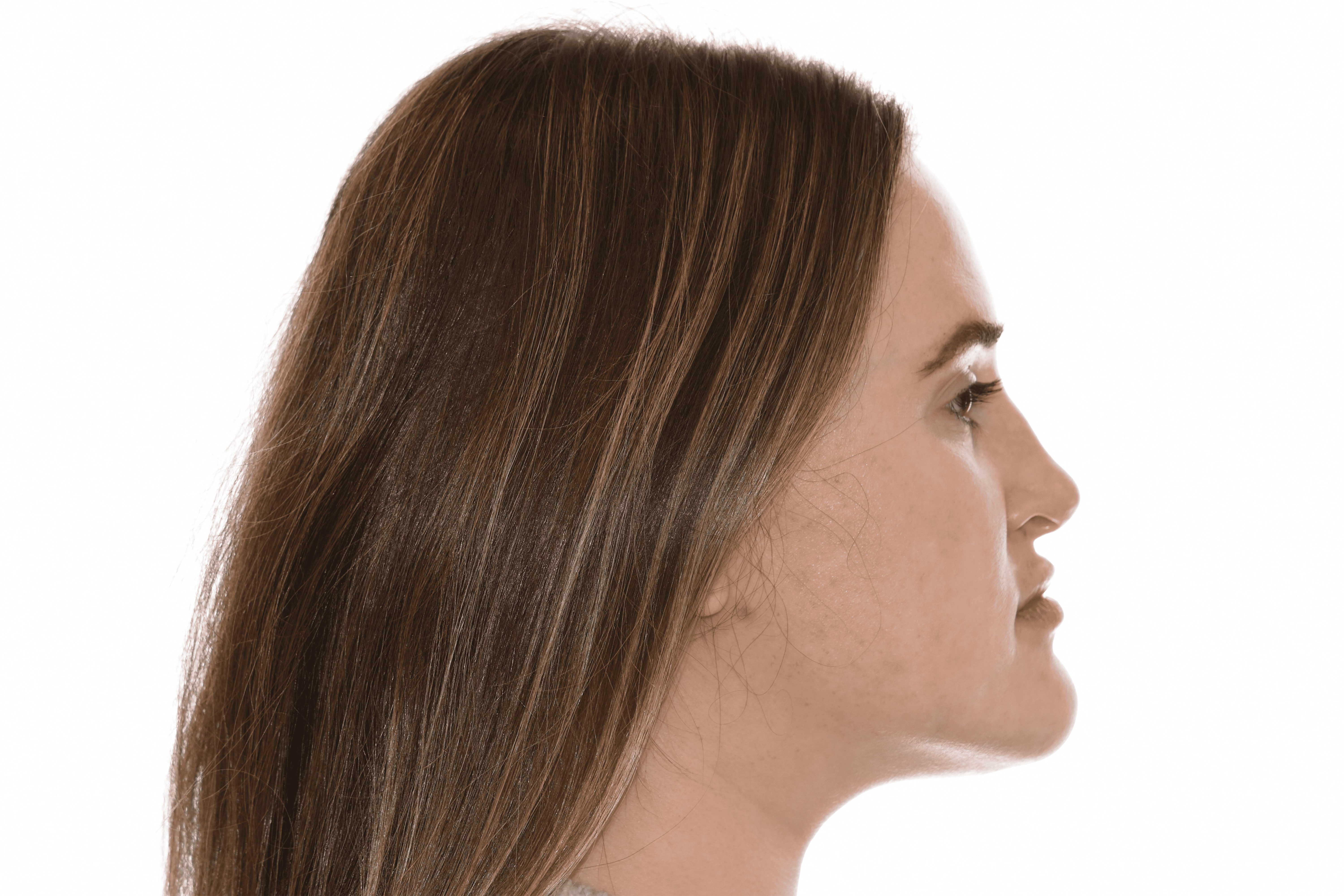
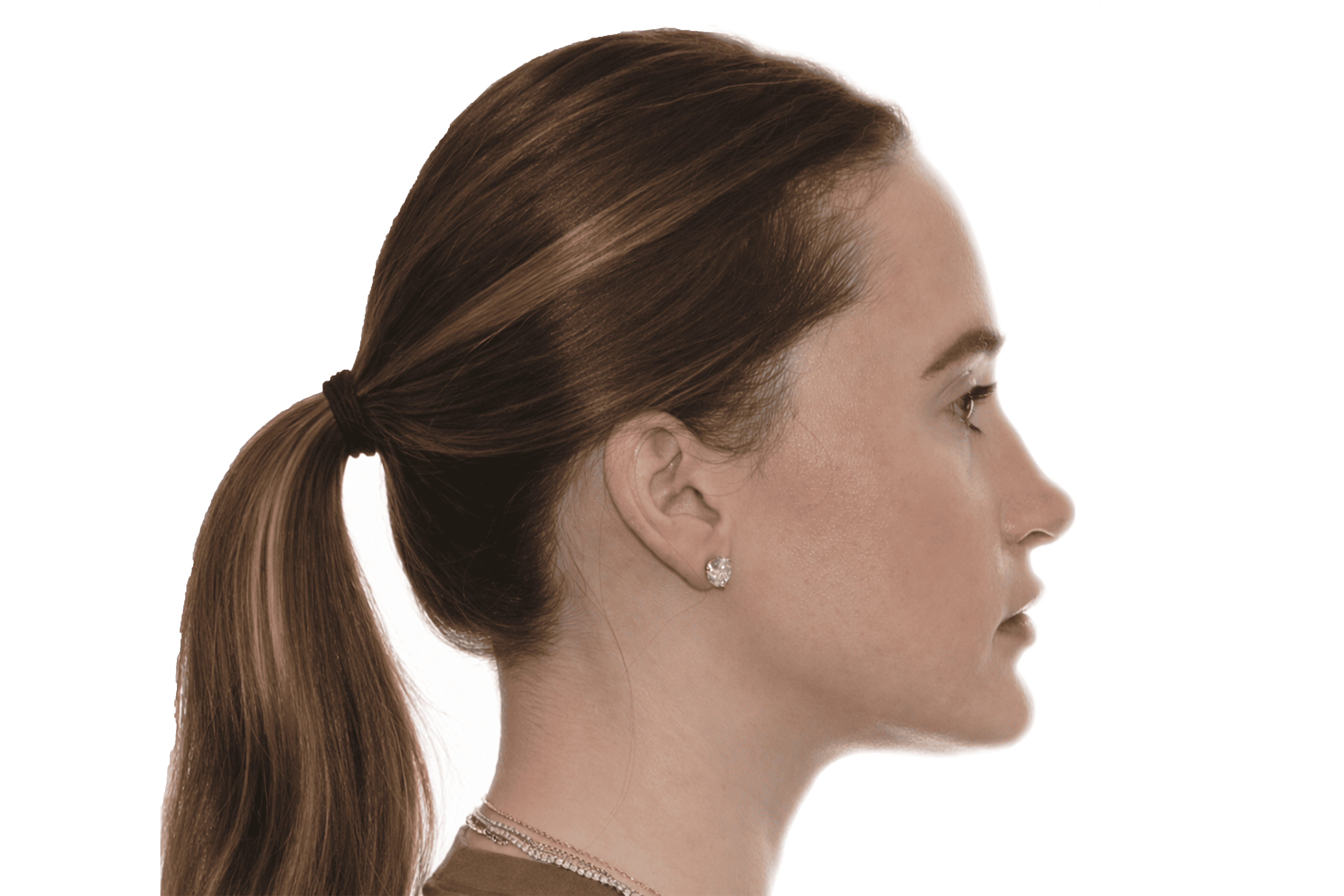

Timeline For Maxillary Distraction
Recovery
Following maxillary distraction, recovery focuses on controlled healing, gradual advancement of the upper jaw, and close monitoring of comfort, progress, and stability.
Week 1
Early Recovery
Week 2–4
Activation Phase
Month 1–3
Consolidation & Device Removal
Month 3–12
Final Results
What To Expect After Maxillary Distraction
Final aesthetic and functional results appear gradually as the bone consolidates and soft tissues adapt.

Risks & Possible Complications

An experienced team and close follow-up care help maximize the likelihood of optimal outcomes.
Maxillary Distraction Osteogenesis requires precise surgical judgment, specialized training, and familiarity with the anatomical and functional nuances of this procedure. Outcomes depend heavily on the surgeon's experience, technique, and ability to anticipate and manage the full range of possible findings. Surgeons who perform this procedure regularly are typically better positioned to achieve consistent, safe results.
In a private practice setting, care is often more streamlined and consistent, with the same surgeon closely involved in evaluation, treatment planning, surgery, and follow-up.
In academic or hospital-based settings, care may involve a larger team that can include residents or fellows participating under supervision. Both models can provide excellent care, but the structure and continuity of treatment may feel different. At our center, your care is delivered directly by your surgeon — not by residents, fellows, or surgeons in training.
It is important to understand who is performing each part of your procedure. In some settings, the attending surgeon performs all critical portions, while in others trainees may assist or participate under supervision. This should be discussed clearly in advance. At our center, your surgeon performs all critical portions of the procedure.
Depending on the treatment setting, residents, fellows, or students may be involved in aspects of care. Patients should feel comfortable asking about their role and how care is supervised.
At our center, there are no residents or fellows involved in performing surgery.
Post-operative follow-up is an important part of surgical care. At our center, your surgeon personally follows you through recovery and any additional visits required.
At our center, your surgeon is directly involved at every stage — from the initial consultation and treatment planning through surgery, post-operative visits, and long-term follow-up.
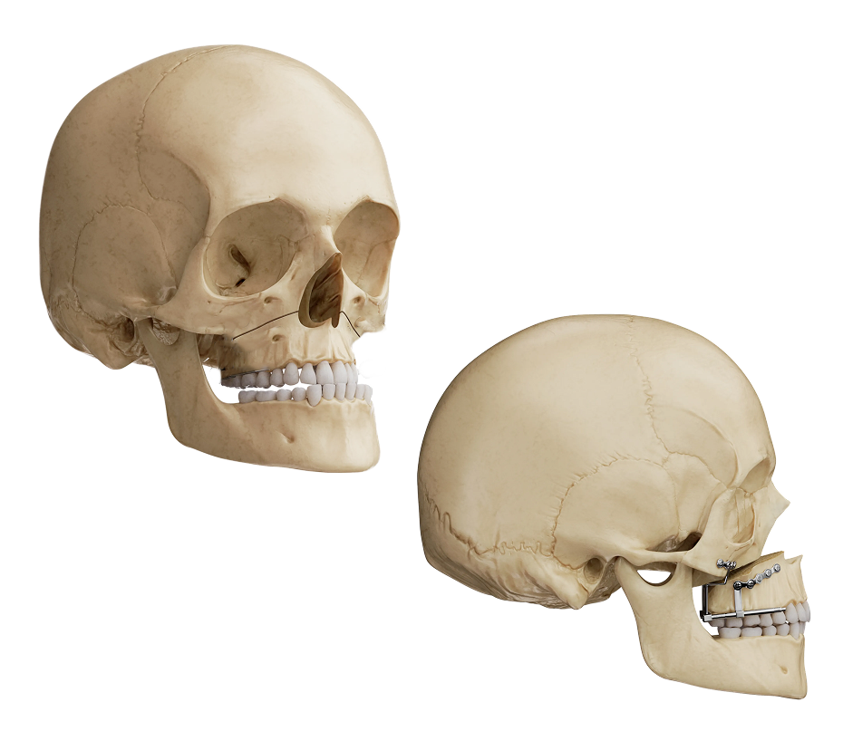
Maxillary distraction osteogenesis is a surgical technique that gradually advances the upper jaw — the maxilla — by making controlled bone cuts and then using a distraction device to slowly move the jaw forward over a period of weeks. New bone forms behind the advancing jaw, allowing the midface to be moved farther forward than is typically achievable with standard Le Fort I or Le Fort III jaw surgery alone.
Maxillary distraction is typically used when the degree of upper jaw advancement required is too large to achieve safely in a single acute movement — generally when movements of approximately 10 mm are needed. This is most commonly seen in patients with cleft lip and palate whose upper jaw is significantly retrusive, and in patients with syndromic midface deficiency such as Crouzon or Apert syndrome.
Large acute advancements carry a higher risk of relapse and can place tension on the soft tissues and airway. Distraction allows the soft tissue envelope to expand gradually alongside the bone, reducing relapse rates and enabling greater movements. It also generates new bone in the gap, which can be advantageous in patients with prior bone grafts or cleft-related bone deficiency.
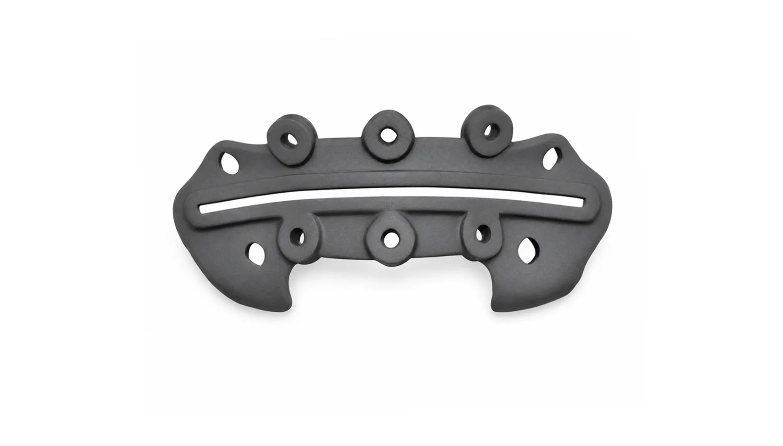
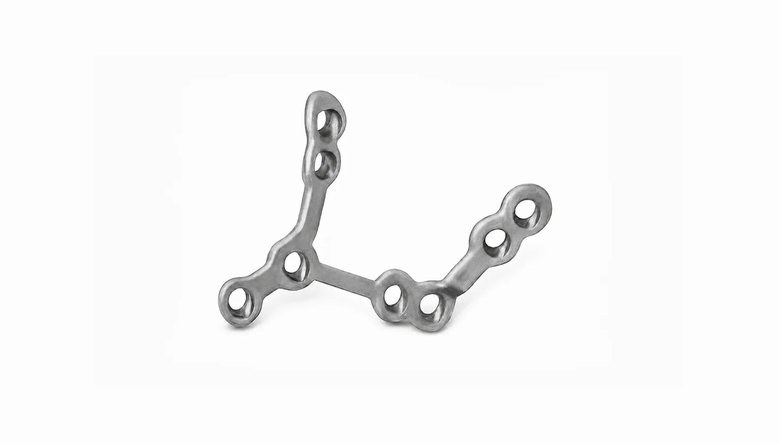
Both internal and external distraction devices are used depending on the patient's anatomy, the degree of advancement required, and the surgeon's preference. Internal devices are placed through incisions inside the mouth and are less visible, while external halo-type devices allow greater control over the direction of movement and are sometimes preferred for very large advancements. The device selection is discussed during treatment planning.
Like mandibular distraction, the treatment occurs in three phases: a latency phase of several days after surgery before distraction begins, an active distraction phase during which the device is turned incrementally each day over several weeks, and a consolidation phase during which the device remains in place while new bone matures — typically six to eight weeks before device removal.
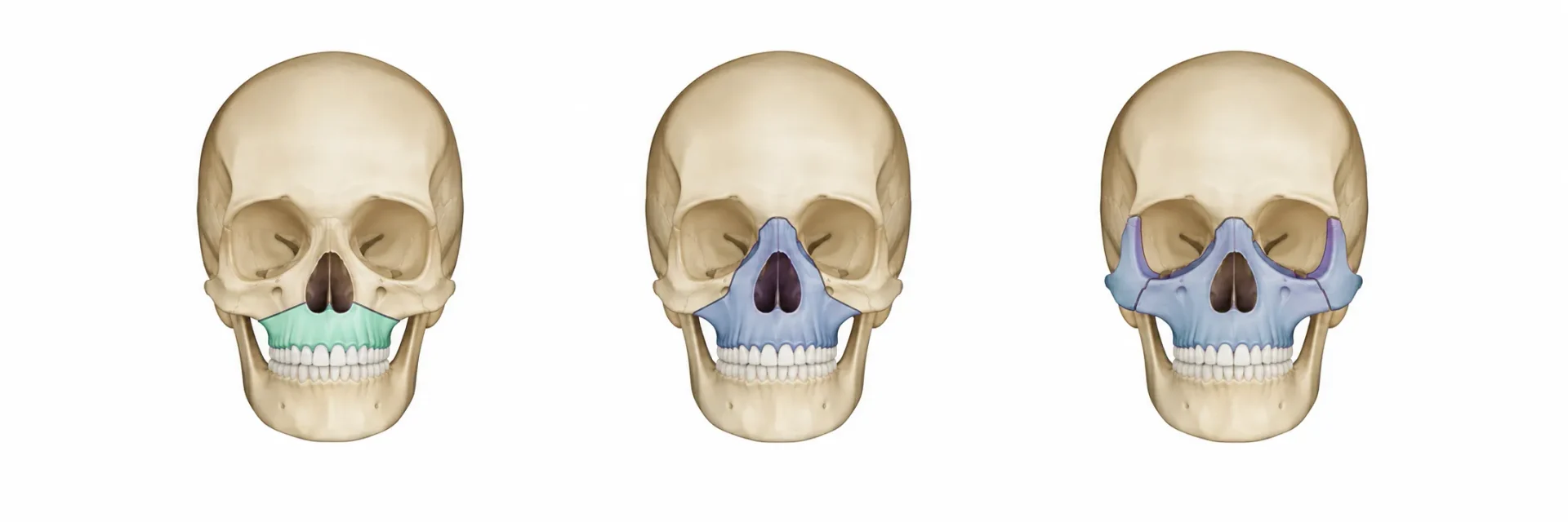
The initial procedure involves making bone cuts through the upper jaw — typically at the Le Fort I or Le Fort III level depending on the extent of midface involvement — and attaching the distraction device. The procedure is performed under general anesthesia. Device placement may require both intraoral and external incisions depending on the device type.
Families are trained before discharge on the activation protocol, which involves turning the distraction device a prescribed number of turns each day. Activation is typically performed at home, with scheduled follow-up visits to monitor jaw position, device stability, and healing. Close attention to the activation protocol is important for achieving the planned result.
Once the planned advancement is achieved, the device is left in place during the consolidation phase while the new bone hardens. Device removal is then performed in a separate surgical procedure under general anesthesia. Additional orthodontic and surgical refinement may be planned after consolidation depending on the patient's needs.
Yes. We regularly care for patients and families traveling from outside the region for maxillary distraction osteogenesis.
Maxillary distraction involves an extended active treatment phase, and the local stay is more involved than a standard jaw surgery procedure. Depending on the case, families may remain locally through the full active distraction phase to allow close monitoring by the surgical team, or may travel home after initial surgery and activation training and return at key intervals. The consolidation phase follow-up may incorporate telemedicine visits for appropriate patients. Device removal requires a return to the center once consolidation is confirmed on imaging. The full plan is discussed with families in detail before surgery so that travel and scheduling can be arranged well in advance.
Yes. Follow-up after maxillary distraction includes monitoring of the advanced jaw position, bite development, bone consolidation, and the need for any future orthodontic or surgical refinement. Some visits may be conducted via telemedicine while others require an in-person return. Your surgeon will outline the complete follow-up plan before the family travels home.
The consultation includes clinical and imaging assessment of the midface, review of prior surgical history, and a detailed discussion of the distraction plan, device options, activation protocol, expected timeline, and what the local stay and return visits will involve. Families and patients leave with a thorough understanding of each phase of treatment.