



Fronto-Orbital Advancement
Reshaping the Forehead and Eye Area to Support Healthy Growth and Development
What Is Fronto-Orbital Advancement (FOA)?
What Patients Find Most Helpful to Know About Fronto-Orbital Advancement

Supporting Brain Growth
The primary goal is to protect brain development while improving skull shape.

Timing Matters
Performed early to guide normal brain growth and skull development.

Temporary Swelling Is Expected
Swelling around the forehead and eyes is common and improves steadily over weeks.

Two Surgical Options
An endoscopic or open technique may be performed, based on age and severity.
When Is Fronto-Orbital Advancement Performed?

Improved protection for the brain and eyes
Normalized forehead and orbital contours
Reduced risk of intracranial pressure complications

Enhanced facial symmetry and proportion

Support for healthy neurological development

Long-term stability as the child grows

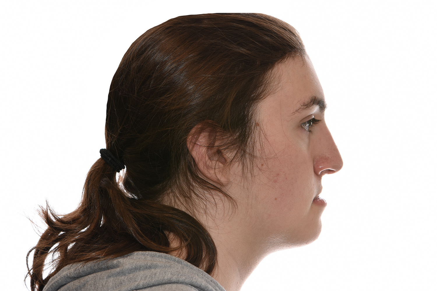
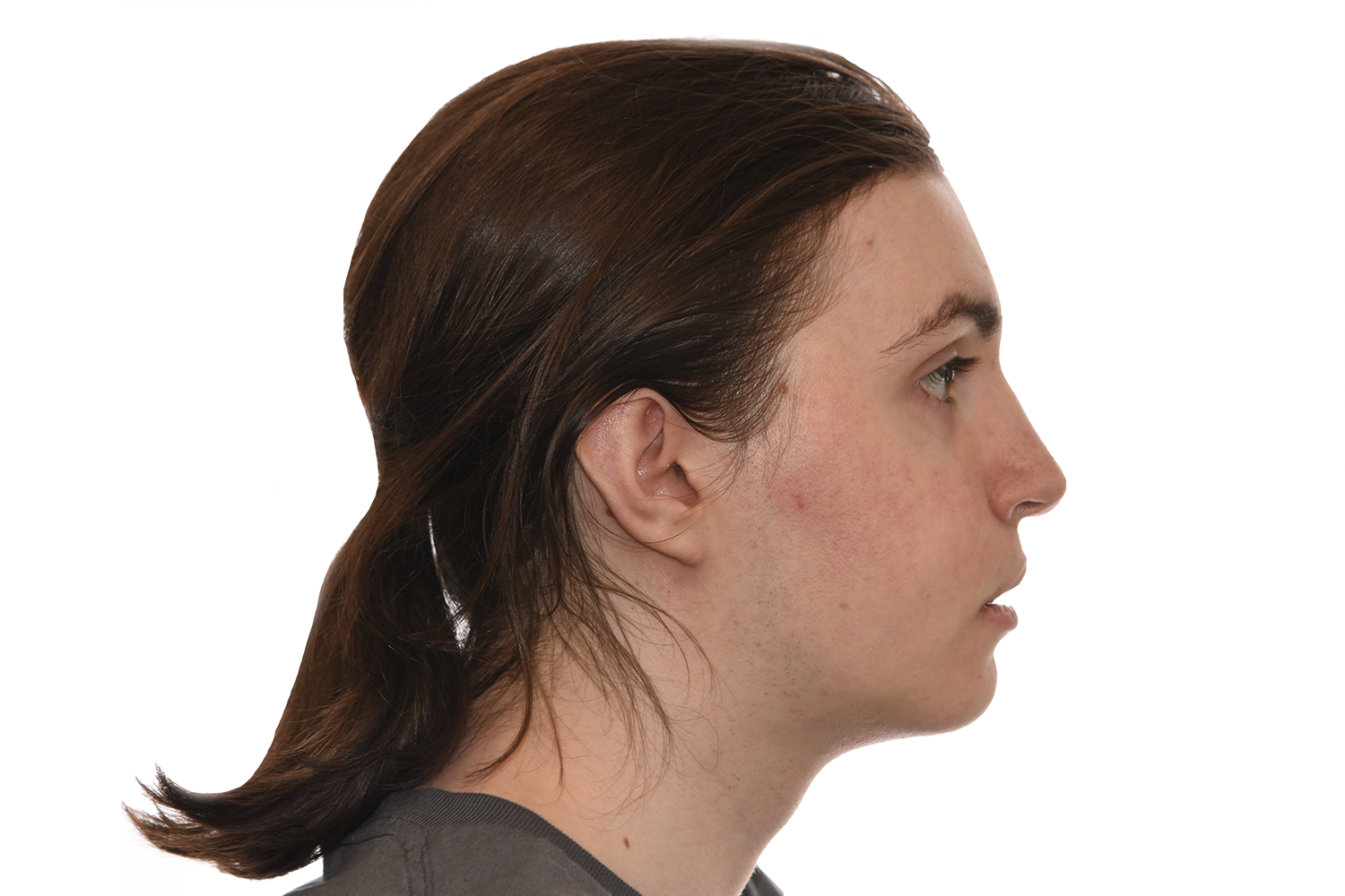
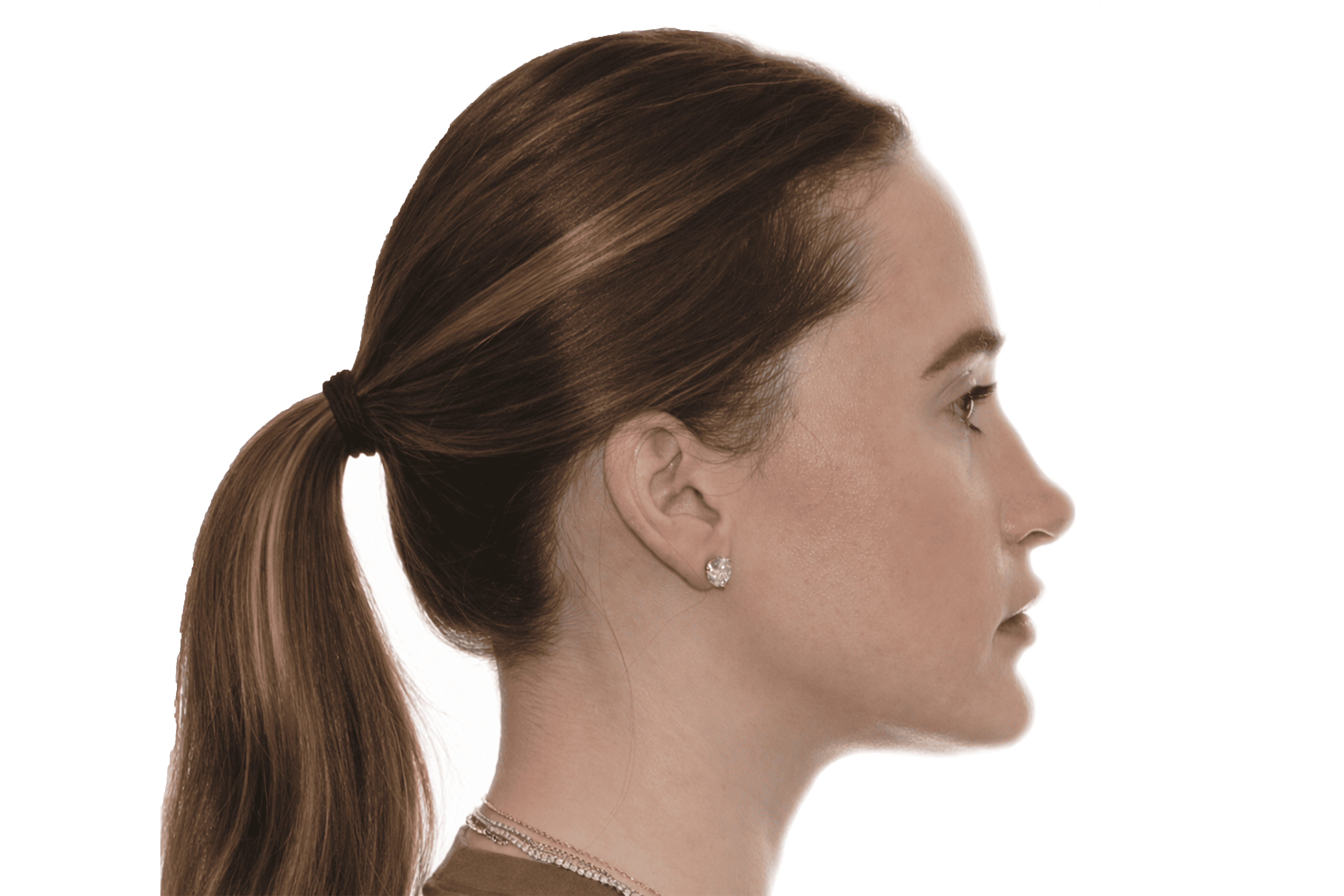
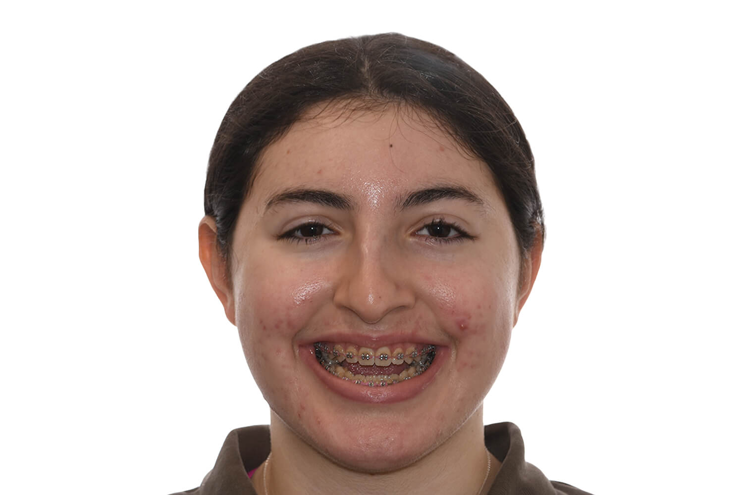
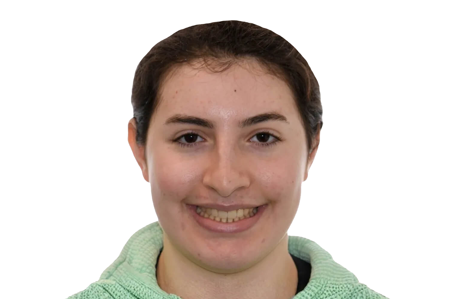
Before & After Gallery

Timeline For Fronto-Orbital Advancement
What To Expect After FOA
Most children recover well and resume normal development with ongoing monitoring.

Risks & Possible Complications

An experienced team and close follow-up care help maximize the likelihood of optimal outcomes.
Frontal Orbital Advancement requires precise surgical judgment, specialized training, and familiarity with the anatomical and functional nuances of this procedure. Outcomes depend heavily on the surgeon's experience, technique, and ability to anticipate and manage the full range of possible findings. Surgeons who perform this procedure regularly are typically better positioned to achieve consistent, safe results.
In a private practice setting, care is often more streamlined and consistent, with the same surgeon closely involved in evaluation, treatment planning, surgery, and follow-up.
In academic or hospital-based settings, care may involve a larger team that can include residents or fellows participating under supervision. Both models can provide excellent care, but the structure and continuity of treatment may feel different. At our center, your care is delivered directly by your surgeon — not by residents, fellows, or surgeons in training.
It is important to understand who is performing each part of your procedure. In some settings, the attending surgeon performs all critical portions, while in others trainees may assist or participate under supervision. This should be discussed clearly in advance. At our center, your surgeon performs all critical portions of the procedure.
Depending on the treatment setting, residents, fellows, or students may be involved in aspects of care. Patients should feel comfortable asking about their role and how care is supervised.
At our center, there are no residents or fellows involved in performing surgery.
Post-operative follow-up is an important part of surgical care. At our center, your surgeon personally follows you through recovery and any additional visits required.
At our center, your surgeon is directly involved at every stage — from the initial consultation and treatment planning through surgery, post-operative visits, and long-term follow-up.
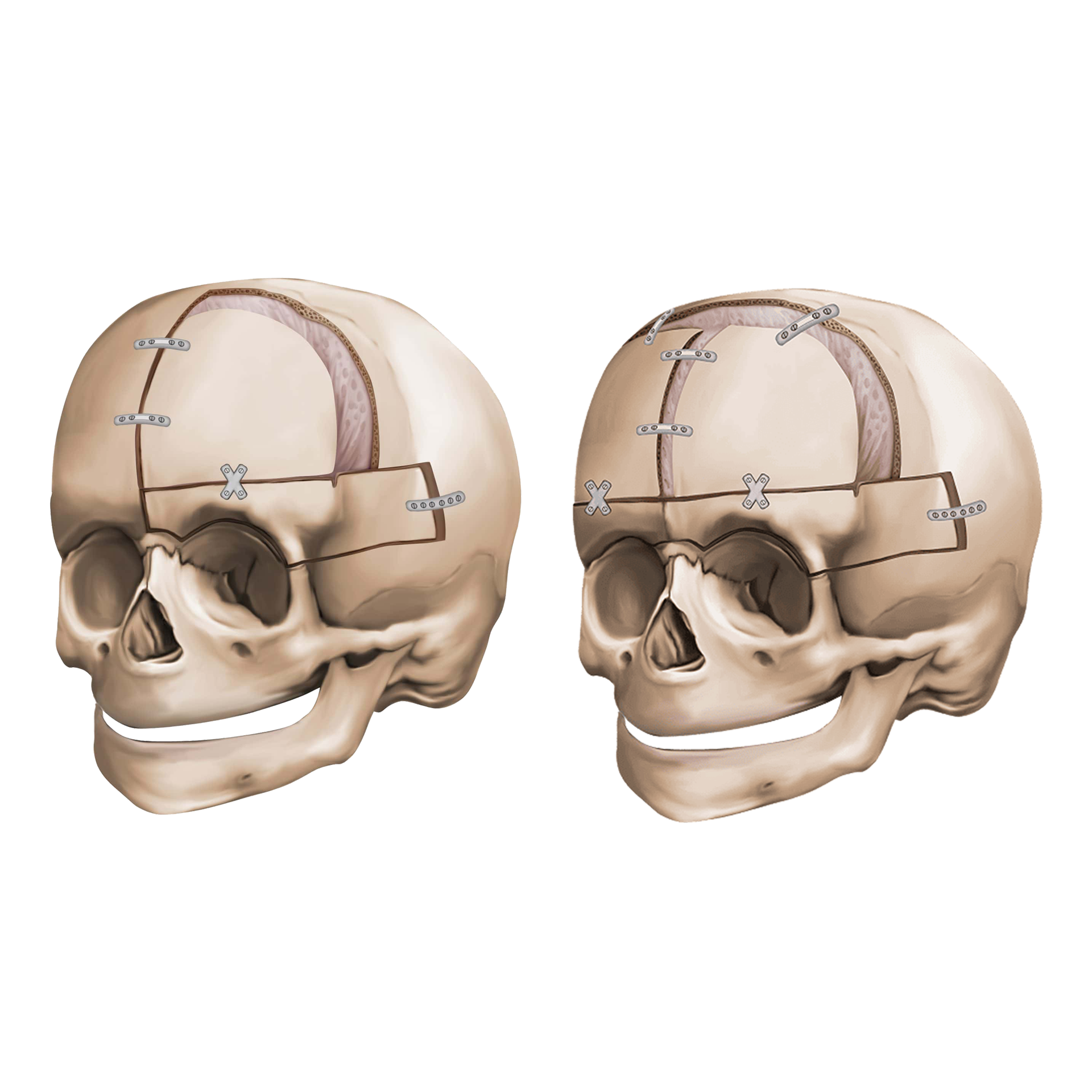
Frontal orbital advancement is a surgical procedure that moves the forehead and the upper rim of the eye sockets — the supraorbital bar — forward to correct the recessed forehead and prominent eyes seen in patients with metopic or coronal craniosynostosis, and in syndromic craniosynostosis conditions. It is typically performed in combination with frontal cranial vault remodeling to reshape the forehead and improve head shape simultaneously.
When the forehead or supraorbital region is restricted by premature suture fusion, the forehead may be flat or recessed and the eyes may appear prominent due to shallow orbits. Frontal orbital advancement moves these structures forward, creating a more normal forehead contour, improving orbital depth, and relieving pressure on the brain from restricted skull growth.
The traditional open approach involves reshaping and repositioning the frontal bone and supraorbital bar through a coronal scalp incision and securing them with plates or resorbable fixation. A distraction-assisted approach is also used in some patients, particularly when larger advancements are needed or when gradual movement is preferred to reduce the risk of relapse and accommodate soft tissue expansion. The appropriate technique is determined by the patient's anatomy, age, and the degree of advancement required.
Endoscopic techniques are used for early suture release in young infants, but frontal orbital advancement as a distinct procedure is typically performed using open or distraction-assisted approaches. The choice depends on the patient's age and the degree of frontal and orbital retrusion requiring correction.
The procedure involves the bones of the skull immediately adjacent to the brain and typically requires collaboration between the craniofacial surgeon and a neurosurgeon. The dura — the covering of the brain — is carefully protected throughout the procedure. The intracranial proximity of this surgery is an important reason why the post-operative monitoring period is more extended than for purely facial jaw procedures.
Frontal orbital advancement is performed under general anesthesia through a coronal scalp incision. The neurosurgical and craniofacial teams work together to carefully mobilize the frontal bone and supraorbital bar, advance them to the planned position, and secure them with titanium or resorbable plates and fixation. A blood transfusion is commonly required. The procedure typically takes three to five hours.
Significant swelling of the forehead and around the eyes — including closure of the eyelids — is expected and peaks in the first two to three days before gradually resolving. Children are closely monitored in the hospital during this period. Most children are hospitalized for three to five days and return to normal activity over several weeks, with full bone healing occurring over months.
When distraction is used, the initial surgery is followed by a period of daily activation during which the device gradually advances the frontal and orbital bones. Families are trained on device activation before discharge. Device removal is performed in a second procedure under general anesthesia after consolidation is complete.
Yes. We regularly care for families traveling from outside the region for frontal orbital advancement surgery.
Frontal orbital advancement involves the skull adjacent to the brain, and the local stay reflects the importance of close post-operative monitoring. After the open procedure, families should plan for a total local stay of approximately two to three weeks from the time of surgery. The hospital stay is three to five days, with the remaining time needed to monitor intracranial pressure indicators, orbital swelling, healing, and overall recovery before travel home. For distraction-assisted frontal orbital advancement, families must remain locally through the active distraction phase and return for device removal once consolidation is confirmed. The full plan is discussed in detail before surgery.
Yes, and this distinction is important after frontal orbital advancement given the intracranial nature of the procedure. Ground travel is generally approved before air travel. Flying is typically cleared at approximately two to three weeks after the open procedure, once the team is satisfied with healing and intracranial monitoring. Your surgeon will advise on both clearances based on your child's recovery.
Yes. Long-term monitoring of skull growth, intracranial pressure, orbital development, and vision is an important part of post-operative care after frontal orbital advancement. Some follow-up may be conducted via telemedicine while key assessment visits will require an in-person return. Your surgeon will outline the full follow-up plan before the family travels home.
The consultation includes cranial and orbital examination, imaging review, neurosurgical assessment, and a thorough discussion of the procedure, timing, recovery, and what the local stay and long-term follow-up will involve. Families leave with a clear understanding of the surgical plan and what to expect at each stage.