

Young man with maxillary hypoplasia in jacket and tie smiling

Maxillary Hypoplasia

A condition we regularly treat, maxillary hypoplasia is highly correctable with outcomes that improve midface support, bite alignment, and overall balance.

What Is Maxillary Hypoplasia

Understanding Maxillary Retrognathia

Maxillary hypoplasia is a condition in which the upper jaw does not develop to its full size or position. It is commonly seen in patients with a history of cleft lip and palate and can affect both facial balance and the way the teeth fit together. When the upper jaw is underdeveloped, it may cause difficulty with biting, chewing, and speaking. Treatment often involves orthognathic (jaw) surgery to reposition the jaws, improving function, alignment, and overall facial harmony.
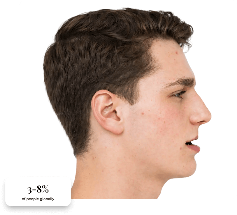
Clinical presentation of maxillary retrognathia, with statistics.

Clinical Presentation

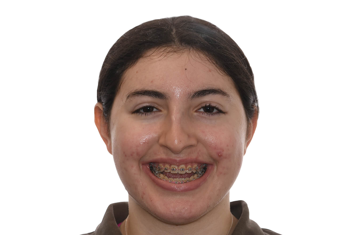
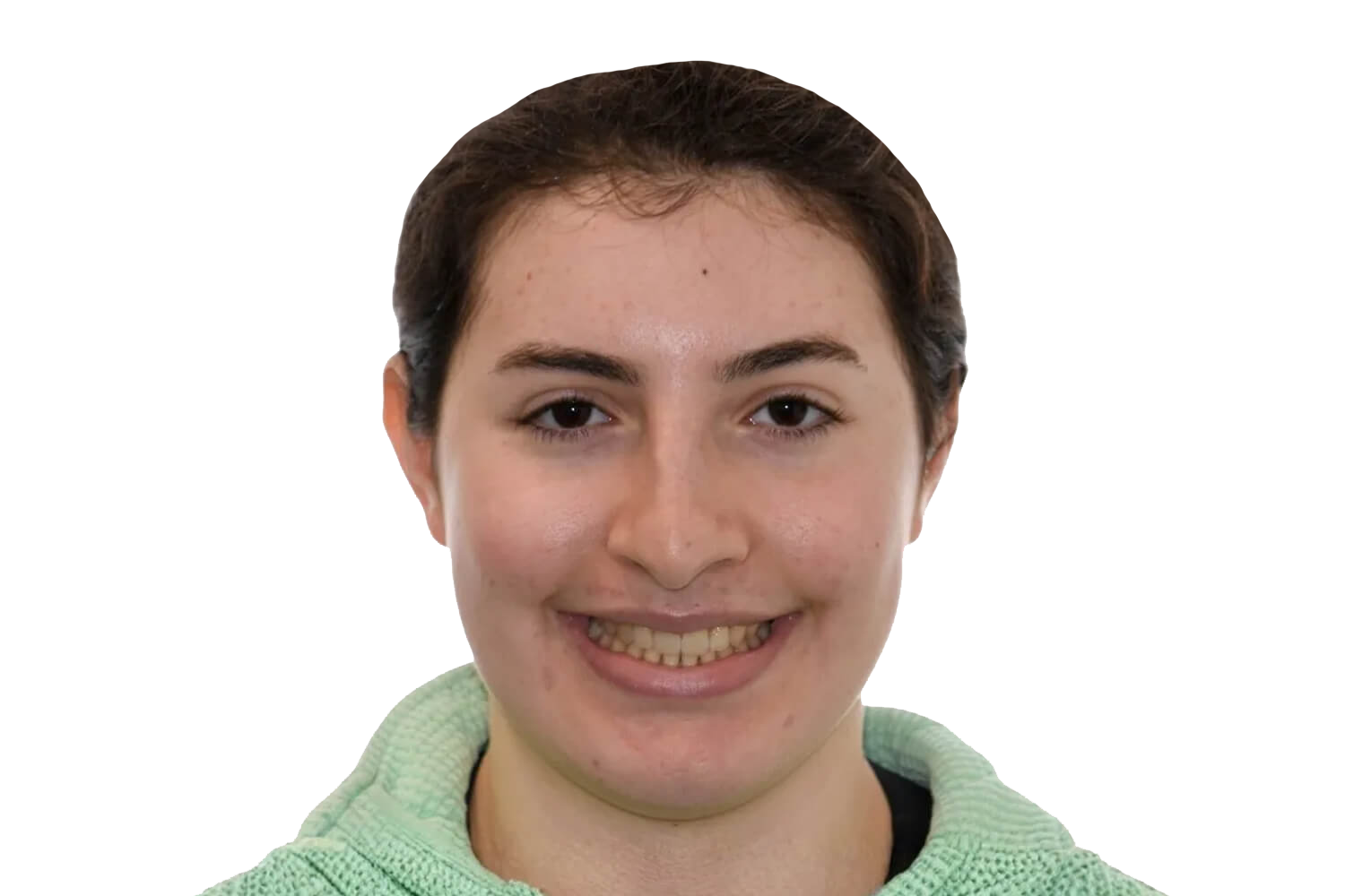
Clinical presentation of maxillary retrognathia, frontal view.
Frontal View

Frontal Analysis

In maxillary hypoplasia, the upper jaw sits farther back than it should, which can affect the projection and balance of the middle of the face. From the front, the midface may appear flatter or less developed compared to the lower jaw.

Key Assessments:

Retruded upper jaw position
Midface deficiency
Flattened facial contours
Reduced upper lip support
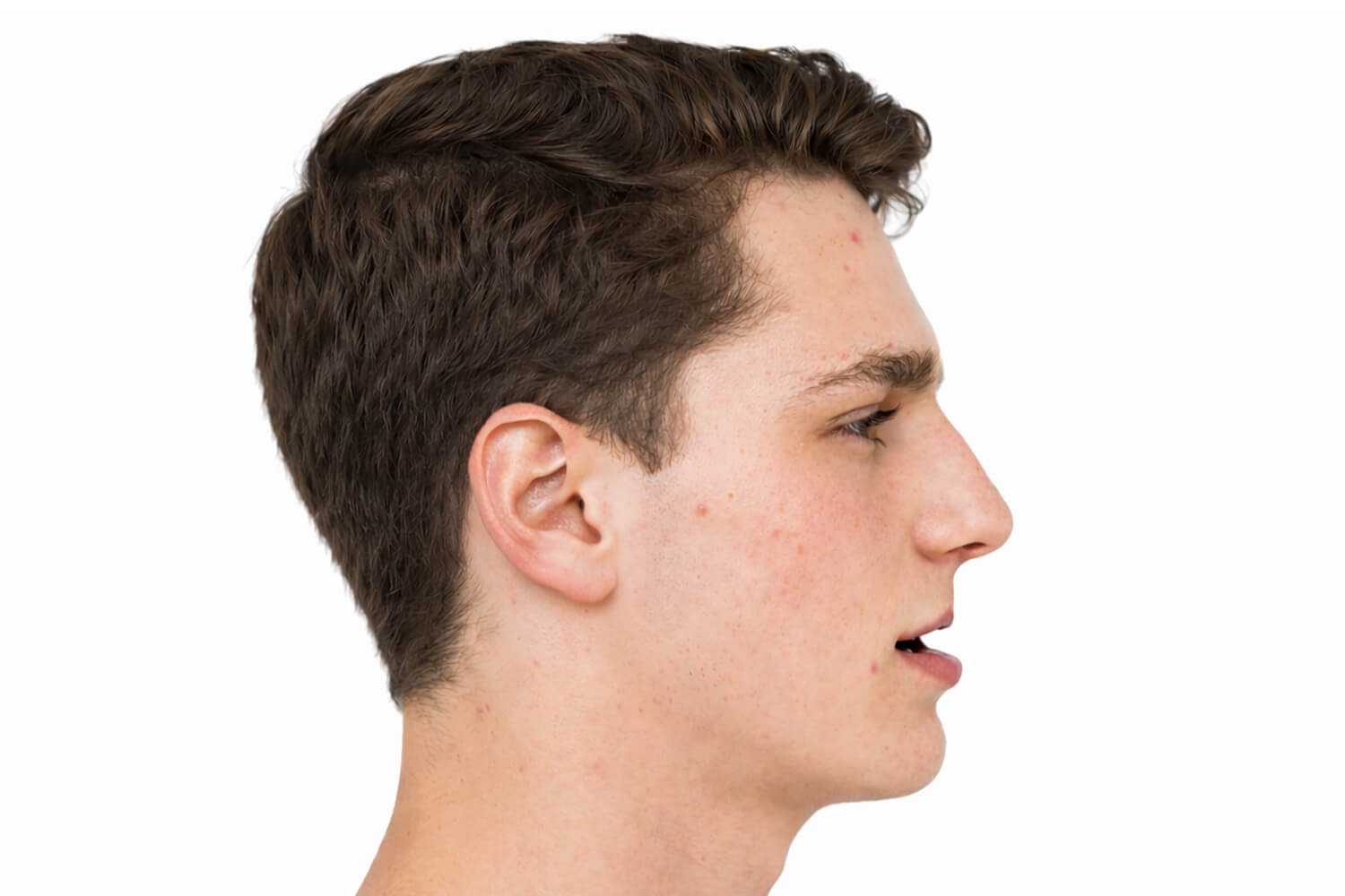
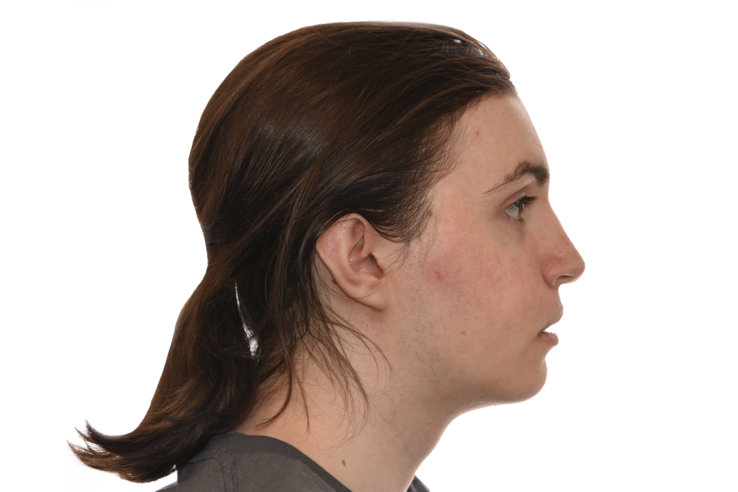
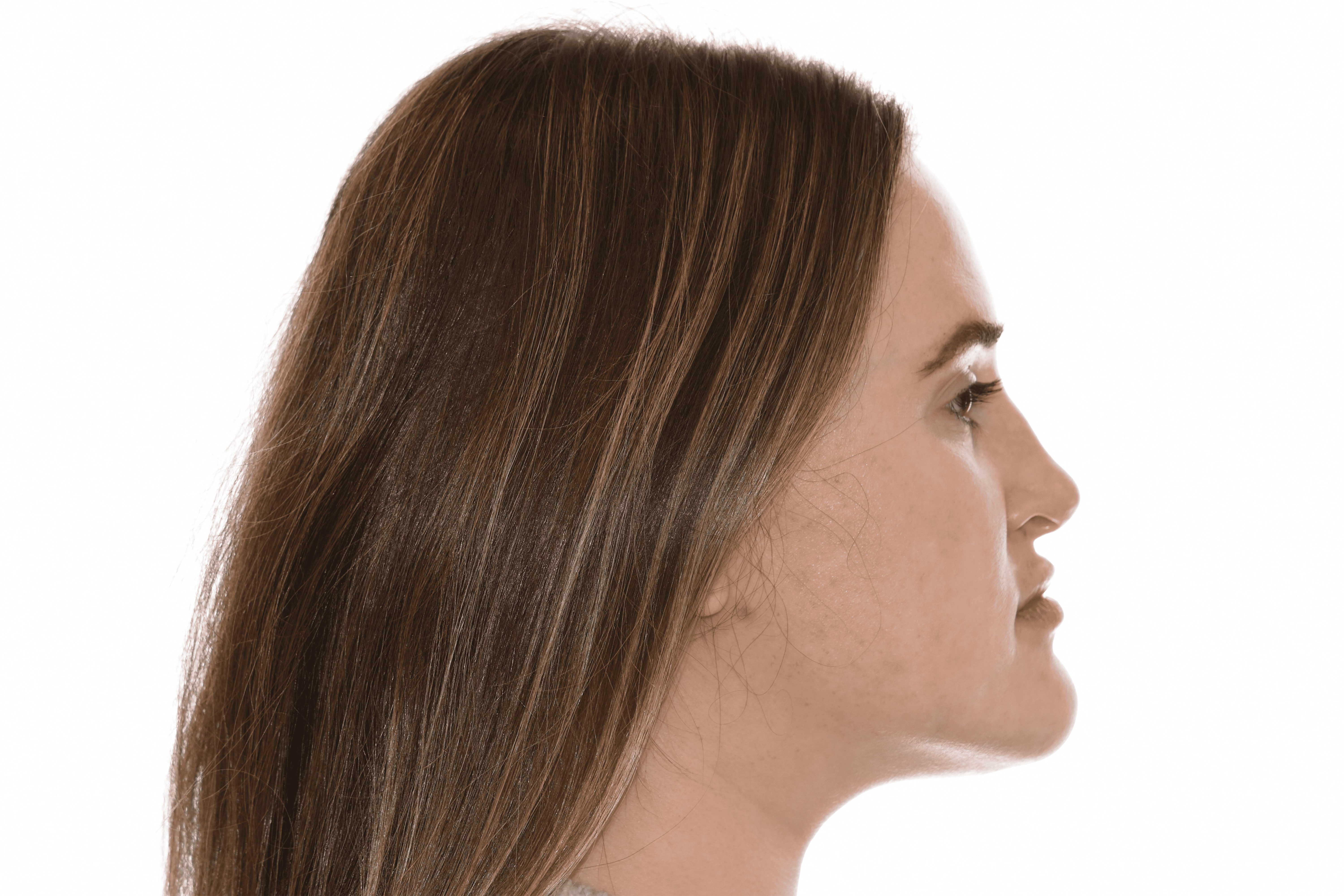
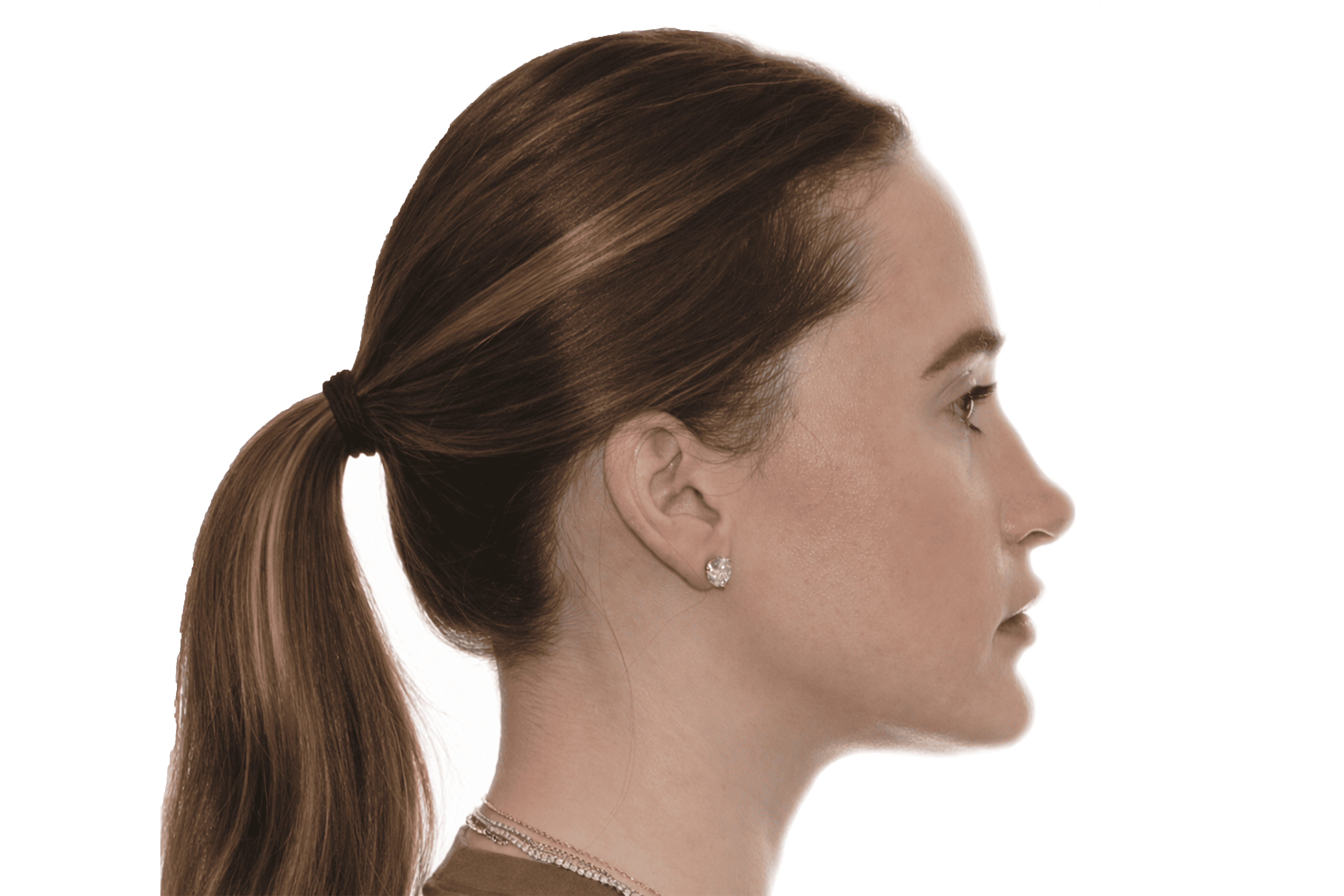
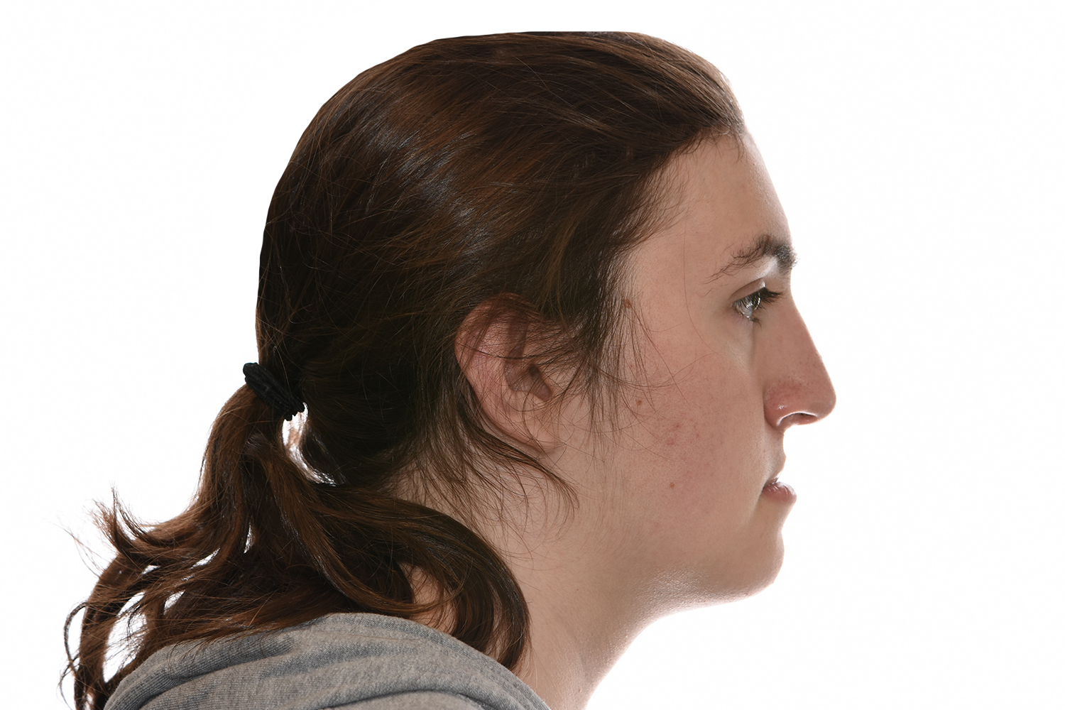
Clinical presentation of maxillary retrognathia, profile view.
Side View

Profile Analysis

From the side, the midface appears set back or underdeveloped, while the lower jaw may look more prominent. The profile often has a concave appearance due to reduced forward projection of the upper jaw.

Key Assessments:

Retruded maxilla
Concave facial profile
Midface under projection
Relative mandibular prominence
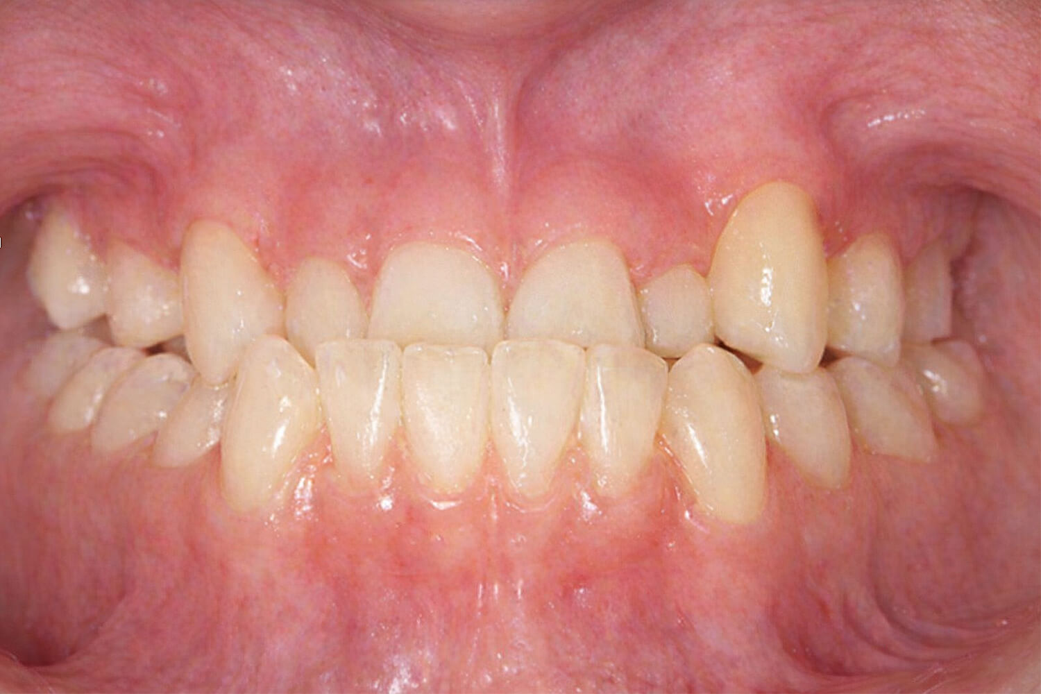
Clinical presentation of maxillary hypoplasia, occlusal view.
Bite View

Occlusal Relationship

Maxillary hypoplasia often results in a bite relationship where the lower teeth are positioned ahead of the upper teeth, commonly reflecting a Class III occlusion.

Key Assessments:

Class III occlusion pattern
Negative overjet relationship
Lower teeth ahead of upper
Dental arch discrepancy

Maxillary Hypoplasia

Common Functional Issues

Icon representing eating and chewing.

Eating

An underdeveloped upper jaw can make it difficult to bite and tear food efficiently with the front teeth.

Icon representing speech.

Speech

Certain sounds may lack clarity due to insufficient upper tooth support for proper tongue positioning.

Icon representing TMJ symptoms.

TMJ

Imbalanced jaw relationships can contribute to uneven joint loading and potential discomfort during function.

Icon representing breathing.

Mouth Breathing

Midface deficiency may affect nasal airflow, encouraging mouth breathing habits over time.

Icon representing nasal congestion.

Nasal Congestion

Reduced midface projection can narrow the nasal airway, impacting airflow and breathing efficiency.

Icon representing physiological factors.

Psychosocial Effects

A flatter midface profile may affect facial balance, influencing how patients perceive their appearance.

Before & After Gallery
Maxillary Hypoplasia

Jaw Surgery Procedures

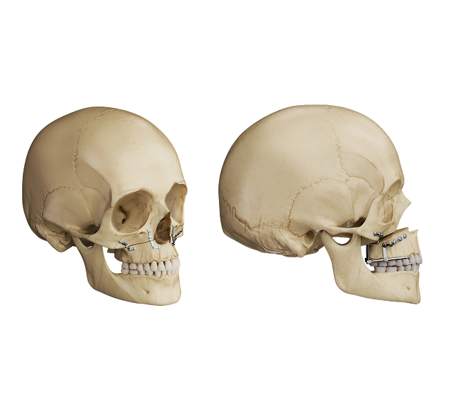
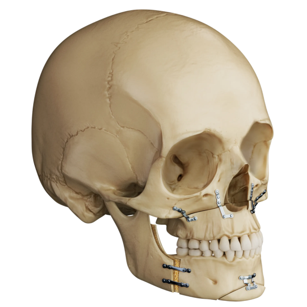
Orthognathic surgery corrects maxillary hypoplasia by repositioning the upper jaw to improve bite function, midfacial support, and balance. Common procedures include Le Fort I osteotomy, BSSO, double jaw surgery, and in severe cleft-related cases, maxillary distraction.

BSSO

BSSO Result Illustration

Le Fort I

Illustration of maxillary distraction at the Le Fort I level.

Double Jaw

Double Jaw Surgery Result Illustration

Life-Changing Benefits

Advancing the upper jaw improves bite alignment, midface support, and breathing efficiency.

Improved bite function
Enhanced midface projection
Better nasal airflow
Improved speech clarity
Balanced facial profile
Increased confidence

Surgical Management of Maxillary Hypoplasia

Upper Jaw

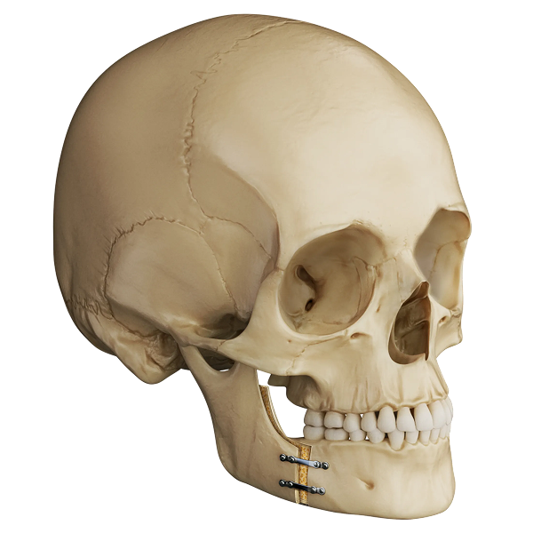
Le Fort I osteotomy corrects maxillary hypoplasia by advancing the upper jaw forward to restore midface projection and correct the concave facial profile. In cleft-related cases, where scar tissue limits conventional advancement, distraction osteogenesis may be recommended to gradually move the jaw into position. The jaw is secured with titanium plates and screws, improving chewing, speech, and facial proportions. Each case is planned with advanced imaging and surgical simulation.

Both Jaws

When maxillary hypoplasia is severe or accompanied by additional skeletal discrepancies, double jaw surgery is recommended. A Le Fort I osteotomy advances and repositions the upper jaw while a bilateral sagittal split osteotomy simultaneously repositions the lower jaw to optimize the bite and facial profile. In cleft-related cases, distraction osteogenesis may be incorporated to achieve the full degree of upper jaw advancement required. Both jaws are secured with titanium plates and screws, improving chewing, speech, breathing, and facial balance.

Presurgical Considerations

Portrait handsome young man.

Growth Completion

Key Assessment Points:
Midface growth complete
Alveolar (cleft) Addressed
Occlusion no longer changing
Serial ceph unchanged
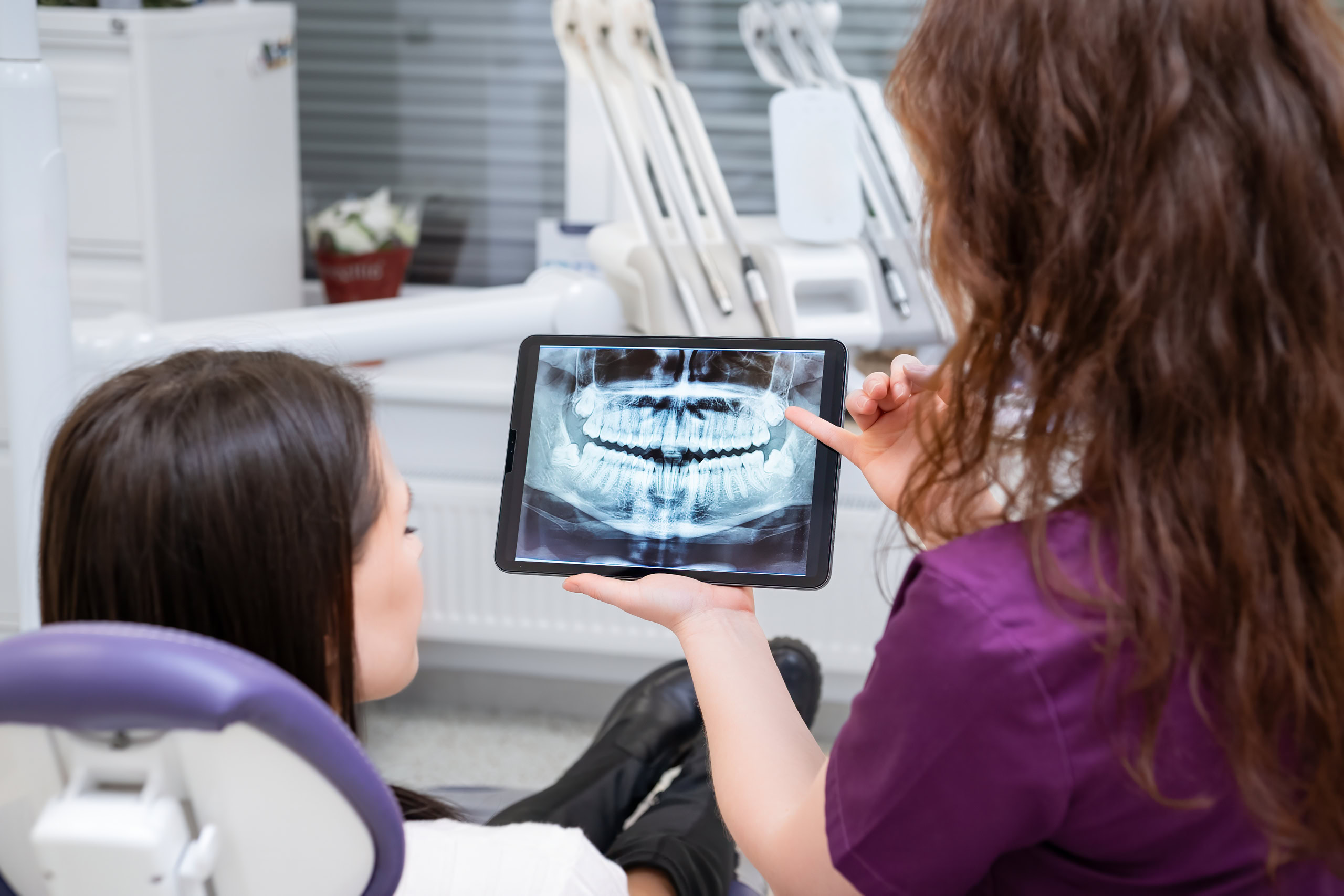
Dentist reviewing X-rays with patient on tablet

Orthodontic Preparation

Key Assessment Points:
Maxillary incisor decompensation
Overjet increased pre-surgery
Upper incisor uprighting
Transverse coordination optimized
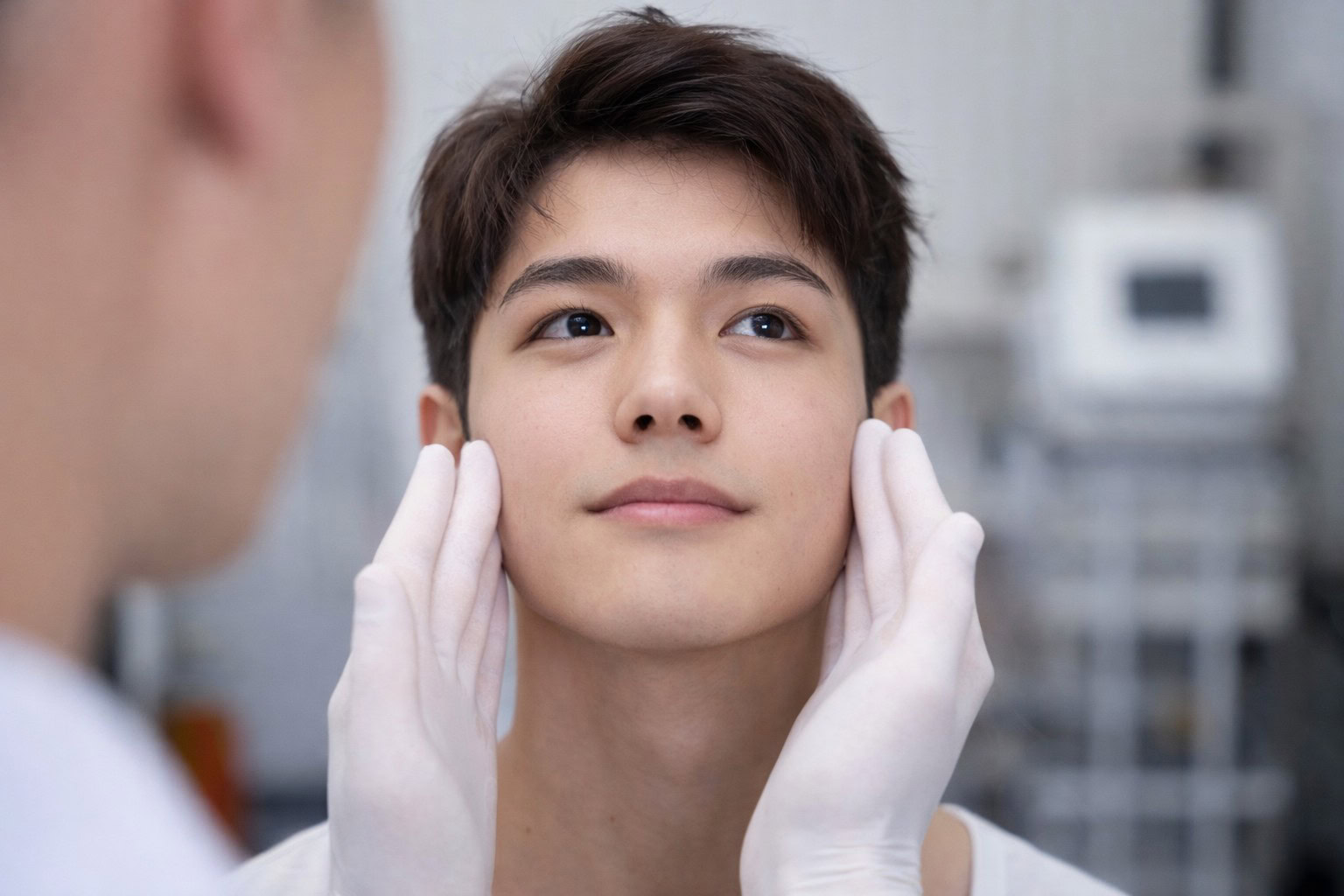
Doctors consultation.

Midface & Cleft Assessment

Key Assessment Points:

Presence of fistula (cleft)
Nasal airway assessment
Incisor bone support (cleft)
Soft tissue profile review

Timeline for Maxillary Hypoplasia

Month 0

Initial Consultation

Comprehensive history, clinical evaluation, 3D scans, X-rays, photographs, and impressions to assess jaw position and bite.

2–4 weeks after initial consultation

Treatment Planning Visit

Once your records are reviewed and the doctor has coordinated with your orthodontist, you’ll return either in person or via telemedicine to review your treatment plan, see prediction images, and go over the steps and timeline ahead.

6–12 months

Orthodontic Preparation (if necessary)

Braces or aligners gradually guide the teeth into proper positions to prepare the jaws for surgical
correction. In select cases, a surgery-first approach may be possible.

3-6 weeks prior to surgery

Final Surgical Planning

Virtual surgical planning determines precise bone movements for optimal symmetry, stability,
and facial balance.

2–4 weeks prior to surgery

Pre-Op Medical Clearance

Medical exams, lab testing, and final instructions ensure the body is fully prepared for surgery.

Typically a one-night hospital stay

Surgery Day

Corrective jaw surgery is performed to reposition the jaws, improve bite function, and enhance facial balance.

Distraction Phase (If Necessary)

Following a one-week healing period, the distraction device is gradually activated over 2 to 3 weeks to advance the upper jaw. A consolidation phase of 1 to 3 months then allows the newly formed bone to mature and stabilize.

3–9 months after surgery

Post-Surgical Orthodontics

Final orthodontic treatment fine-tunes tooth position and bite, optimizing aesthetics, function, and long-term stability.

Experience Matters

“We personally plan your care, perform your surgery, and guide your recovery. No residents, no fellows, no handoffs.

Choosing the right surgical team matters. You should feel informed, comfortable, and confident in every step of the process. At our practice, your care is provided by one of the most highly trained and experienced jaw surgery teams in the country.

When you come to our office, you meet directly with your surgeons. We are the ones who plan your treatment, perform your surgery, see you after surgery, and remain personally available to you throughout your recovery. Unlike larger institutions, your care is not delegated to residents, fellows, or doctors in training. Every critical aspect of your treatment is handled by your surgeons.


Founding Members of the American Society of Orthognathic Surgery

What Our Patients Are Saying
Handsome young man, blue shirt, short hair, smiling
★★★★★

Dr Lee and the team at Northeast Facial are incredible. I was very nervous about my procedure but Dr Lee walked me through it step by step, never once making me feel rushed. Silvanna at the front desk went above and beyond to help me schedule an appointment that worked for me. I would highly recommend NEFOSS!

Stephanie Brown

★★★★★

“I had double jaw surgery done with Dr. Jacob in June 2021. I am extremely happy with my results. No more jaw pain and chewing problems. I also feel much more confident with my new smile. The surgery was definitely well worth it, thanks to Dr. Jacob and his team!”

Amelia Chen

★★★★★

“I would definitely recommend Dr. Jacob and his team at NEFOSS! Dr. Jacob and the team of doctors are very knowledgeable about all aspects of the TMJ process. They are such a nice group of doctors! The office staff is also very helpful and very friendly.”

Tiffany Puopolo

Frequently Asked Questions

Here are some common questions about maxillary retrognathia and available treatment options.

young man with maxillary hypoplasia smiling outside
Take the first step of your journey.

Schedule a private consultation today

Scroll to Top