

Craniosynostosis
Timely treatment guides normal growth, shape, and healthy brain development
What Is Craniosynostosis?
Treatment & Care Pathway

Evaluation & Imaging
Clinical assessment, imaging, and genetic testing are commonly used together to establish the diagnosis and guide treatment decisions.

Surgical Planning
When indicated, surgery is typically performed between 3 and 9 months of age to release fused sutures, reshape the skull, and promote brain development.
Cranial Growth
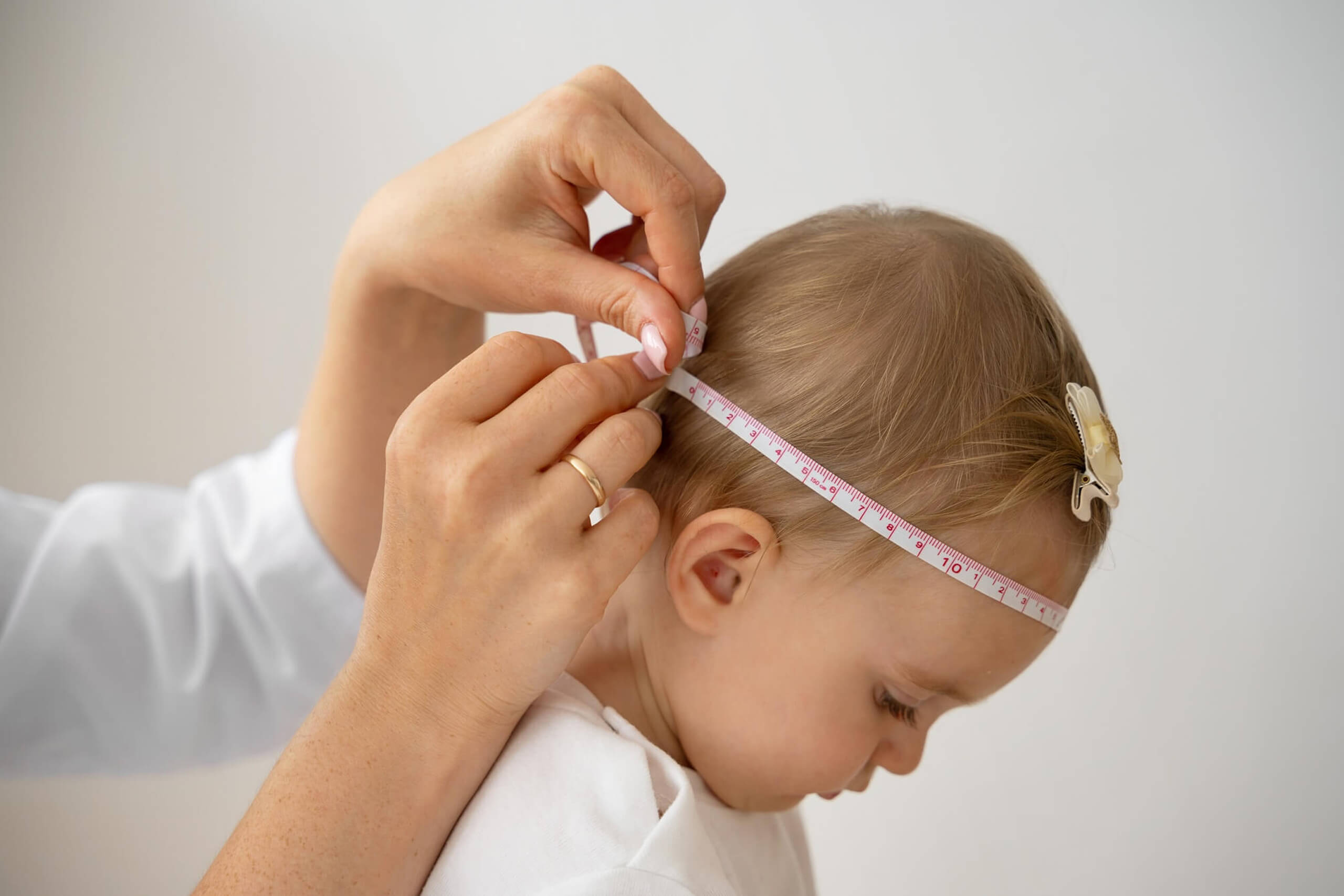
Specialists monitor head growth, brain development, and signs of increased intracranial pressure to determine timing of intervention.

Developmental Care
Ongoing developmental assessments and supportive therapies help address vision, hearing, and developmental needs throughout childhood.
Surgical Treatment Options
Treatment Timeline
Experience Matters
“We personally plan your care, perform your surgery, and guide your recovery. No residents, no fellows, no handoffs.”
Choosing the right surgical team matters. You should feel informed, comfortable, and confident in every step of the process. At our practice, your care is provided by one of the most highly trained and experienced jaw surgery teams in the country.
When you come to our office, you meet directly with your surgeons. We are the ones who plan your treatment, perform your surgery, see you after surgery, and remain personally available to you throughout your recovery. Unlike larger institutions, your care is not delegated to residents, fellows, or doctors in training. Every critical aspect of your treatment is handled by your surgeons.




Founding Members of the American Society of Orthognathic Surgery
What Our Patients Are Saying




Non-Syndromic Craniosynostosis often involves complex surgical, developmental, and functional concerns that require coordinated, specialized management. Outcomes depend heavily on a surgeon's training, clinical judgment, and familiarity with the nuances of this condition. Teams that regularly treat patients with Non-Syndromic Craniosynostosis are typically better equipped to manage both immediate surgical needs and long-term outcomes.
In a private practice setting, care is often more streamlined and consistent, with the same surgeon closely involved in evaluation, treatment planning, surgery, and follow-up.
In academic or hospital-based settings, care may involve a larger team that can include residents or fellows participating under supervision. Both models can provide excellent care, but the structure and continuity of treatment may feel different. At our center, your care is delivered directly by your surgeon — not by residents, fellows, or surgeons in training.
It is important to understand who is performing each part of your treatment. In some settings, the attending surgeon performs all critical portions of the procedure, while in others trainees may assist or participate under supervision. This should be discussed clearly in advance. At our center, your surgeon performs all critical portions of the procedure.
Depending on the treatment setting, residents, fellows, or students may be involved in aspects of care. Patients and families should feel comfortable asking about their role and how care is supervised throughout treatment.
At our center, there are no residents or fellows involved in performing surgery.
Long-term follow-up is an important part of care for many conditions managed at our center. In some practices, post-treatment care may be shared among multiple providers, while in others the surgeon remains closely involved throughout recovery and monitoring. At our center, your surgeon personally follows you throughout recovery and long-term care.
Comprehensive care includes direct surgical involvement in evaluation, treatment planning, surgery, and long-term follow-up. Consistency throughout the process helps maintain continuity of care.
Craniosynostosis is a condition in which one or more of the fibrous joints between the bones of an infant's skull, called sutures, fuse prematurely before the brain has finished growing. This early fusion can restrict normal skull expansion and cause the head to take on a characteristic shape depending on which suture or sutures are involved.
Non-syndromic craniosynostosis refers to premature suture fusion that occurs as an isolated finding, without the other physical features or genetic patterns associated with conditions such as Crouzon, Apert, or Pfeiffer syndrome. It is the most common form of craniosynostosis, accounting for the large majority of cases.
The cause is not fully understood and is likely multifactorial, involving a combination of genetic and environmental influences during fetal development. In most cases, it occurs without a family history and without any identifiable cause. It was not caused by anything either parent did or was exposed to during the pregnancy.
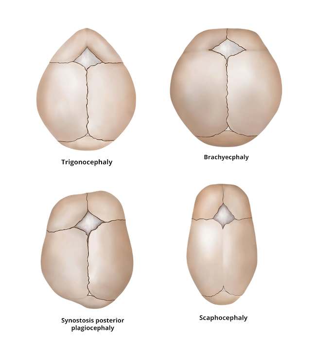
The sagittal suture, which runs along the top of the skull from front to back, is the most commonly fused suture and results in a long, narrow head shape called scaphocephaly. The coronal suture, which runs from ear to ear, is the second most common and can be affected on one side or both. The metopic suture, at the front of the skull, and the lambdoid suture at the back are less commonly involved. Each pattern produces a characteristic head shape.
When a single suture is involved, elevated intracranial pressure is less common than in syndromic forms, but it can occur and should be monitored. The primary concerns in most cases are the shape of the skull and head, and in some cases the development of vision, developmental milestones, and facial symmetry depending on which suture is affected.
In most children with isolated single-suture craniosynostosis who are treated appropriately, development and intelligence are normal. Some children may benefit from early developmental monitoring, particularly if there are concerns about vision or if treatment is delayed.
In most cases where the fusion is confirmed and the skull shape is significantly affected, surgical correction is recommended. Surgery allows the skull to expand appropriately as the brain grows and improves the head shape. The decision to operate depends on the suture involved, the severity of the deformity, and the age of the child at diagnosis.
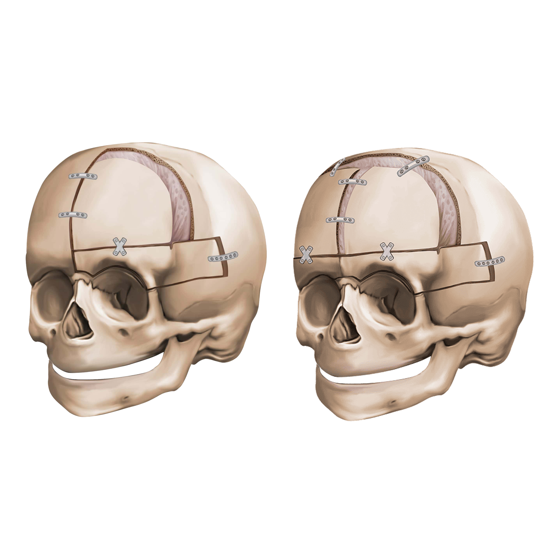
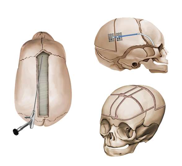
Three main surgical approaches are used depending on the patient's age, the suture involved, and the degree of deformity. Open cranial vault remodeling is the traditional approach, performed through a larger incision with direct reshaping of the skull bones and is applicable across a range of ages. Endoscopic strip craniectomy is a minimally invasive approach performed through small incisions in infants under three to four months of age, removing the fused suture and relying on post-operative helmet therapy to guide skull shape. Spring-assisted cranioplasty is a technique in which small springs are placed at the time of surgery to gradually expand the skull as the brain grows, and is used in selected younger infants for specific suture patterns. The appropriate technique is determined by the child's age, the suture involved, and the severity of the deformity.
For open cranial vault remodeling, surgery is most commonly performed between six and twelve months of age, when the skull bones are still relatively pliable. Endoscopic approaches must be performed in the first few months of life to be effective. Earlier diagnosis and referral improve the range of surgical options available.
Helmet therapy, also called cranial orthosis, is used after endoscopic strip craniectomy to guide skull growth into a more normal shape as the brain expands. It is typically worn for several months following surgery. It is not effective as a standalone treatment for craniosynostosis and does not replace surgical correction.
Craniosynostosis surgery is performed under general anesthesia and involves the skull, so it is taken seriously. Blood transfusion is commonly required for open procedures. Risks include bleeding, infection, and anesthesia-related concerns, and families receive a thorough discussion of these before proceeding. Outcomes are generally very good when surgery is performed by an experienced team.
Yes. After craniosynostosis surgery, children benefit from periodic monitoring of head shape, skull growth, intracranial pressure, vision, and developmental milestones throughout childhood. Most children do extremely well and require no further surgical intervention, though ongoing monitoring helps ensure the best long-term outcome.
Re-fusion of a surgically treated suture can occasionally occur but is uncommon. Regular follow-up allows the care team to monitor for any changes and address them early if needed.
The goal of surgery is to achieve a head shape that is as normal as possible. Results are generally very good, particularly when surgery is performed at the optimal time. Residual asymmetry or shape differences can sometimes be present, and in rare cases additional refinement may be considered as the child grows.
Yes. The large majority of children with non-syndromic craniosynostosis who receive timely surgical treatment go on to live completely normal lives. Early intervention and appropriate follow-up play an important role in achieving the best outcomes.
Yes. We regularly care for families traveling from outside the region for craniosynostosis evaluation and surgical treatment.
The local stay depends on the surgical approach. After open cranial vault remodeling, children are typically hospitalized for three to five days. Once discharged, out-of-town families should plan to remain locally for a total of approximately two to three weeks from the time of surgery, as the early post-operative period involves genuine clinical monitoring of healing, intracranial pressure, and overall recovery before travel is considered safe. After endoscopic strip craniectomy, recovery is faster and hospital stays are shorter, typically one to two nights. Out-of-town families undergoing the endoscopic approach should plan for approximately one week locally, primarily to attend their first post-operative visit where the surgeon confirms healing and initiates helmet fitting before clearing travel home. All timelines depend on the child's age, the procedure performed, and recovery progress, and your surgeon will give you a specific plan well before the procedure.
Yes. Ground travel is generally permitted before air travel, and the distinction is most relevant after open cranial vault procedures. For open remodeling, the intracranial component means that flying clearance typically comes somewhat later than ground travel clearance. For the endoscopic approach, both are generally approved within the first week after the post-operative visit confirms recovery is on track. Your surgeon will advise on both based on the procedure performed and your child's recovery.
Evaluation at our center includes detailed examination of the skull shape, imaging to confirm which sutures are involved and the degree of fusion, assessment of intracranial pressure indicators, and a thorough discussion of surgical options, timing, and what recovery involves. Families leave with a clear understanding of their child's diagnosis, the recommended approach, and what the local stay and follow-up plan will look like.